Category Archives: Photography

Poinsettia flowers part three: the female parts. Stigmas, styles, and ovaries ahoy!

A closeup of a poinsettia flower cluster from directly above it. Many people confuse the bracts (red leaves) with the petals of flowers; they're quite different.   This macro shot shows a number of bracts (colored leaves associated with a flower) surrounding the flowers (green, red, and yellow structures) and extending out of the frame.  The flowers themselves are called cyanthia; the green tissue surrounding each flower is an involucre, a cluster of bracts (leaves) fused into a cup-shaped structure that contains multiple male flowers and one female flower within it.  Emerging from the involucre you can see red filaments supporting yellow anthers on the male flowers; a single female flower should be emerging from the center of each flower.  The bright yellow, liquid-filled structures attached to the involucre are nectar glands filled with yummy sweet nectar to attract pollinators; on less-developed flowers they look like little light-green lips. (Marc C. Perkins)
A closeup of a poinsettia flower cluster from directly above.

[This is the third in a series of three posts. The first post is: Poinsettias up close: where are the flowers? and the second post is: Poinsettia flower closeups take two: getting even closer]

In my previous two posts on poinsettia (Euphorbia pulcherrima) flowers I showed that the flowers are much smaller than most folks think (the large red structures are bracts, not petals) and was able to get some closeups of the inflorescences illustrating their anatomy.

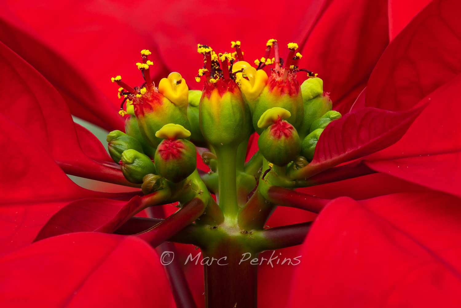
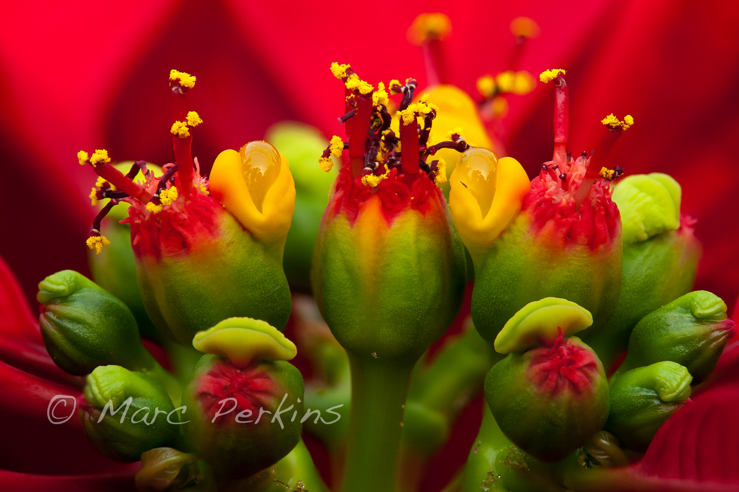
In case you’ve forgotten, the inflorescences of poinsettias are not your typical boring flower: multiple male flowers and a single female flower are surrounded by a sphere of fused bracts called an involucre, out of which the flowers emerge. The involucres often have nectar glands on them, which look like two green or yellow lips.

A closeup of a poinsettia flower cluster from the side.  Many people confuse the bracts (red leaves) with the actual flowers; they're quite different.   This macro shot shows multiple pseudanthia (flowering structures) surrounded by a sea of red bracts (colored leaves associated with a flower).  The flowers themselves are called cyanthia; the green tissue surrounding each flower is an involucre, a cluster of bracts (leaves) fused into a cup-shaped structure that contains multiple male flowers and one female flower within it.  Emerging from the involucre you can see red filaments supporting yellow anthers on the male flowers, and you can even see some of the pollen grains.  Also emerging from each involucre you can see a number of dark-purple structures supported by short stalks (that are about a tenth of the height of the filaments; I'm not sure what these are).   The bright yellow, liquid-filled structures attached to the involucre are nectar glands filled with nectar to attract pollinators. (Marc C. Perkins)
A closeup of a poinsettia flower cluster from the side. The involucre are the large green structures tipped with red, with male flowers emerging from them.

Frustratingly, when I took that first set of pictures I couldn’t find any female flowers on the plants I had. I’ve since learned that this is because the female flowers only develop and extend out of the involucre after the male flowers have matured and produced their pollen (reducing the likelihood of self pollination).  So, I waited, and yesterday finally got a chance to photograph the female flowers up close.

Here’s what a female flower looks like coming out of the involucre:

A single female poinsettia flower with stigma, style, and ovary can be seen emerging from its involucre (cluster of bracts fused together).  The involucre also has a nectar gland emerging from it (that look like two lips).  The entire inflorescence (involcure and female flower) is called a cyanthium.  The red leaves surrounding the inflorescences (mostly out of focus, except for the one next to the female flower) are bracts.  The scale bar is 5mm long. (Marc C. Perkins)
A single female poinsettia flower with stigma, style, and ovary can be seen emerging from its involucre. The scale bar is 5mm long.

There you can see the divided stigma (where pollen need to be deposited if they are to fertilize the flower’s eggs), the swollen ovary just barely protruding from the involucre, and the style connecting those two.

For more context, we can see that the maturing female flower is next to cyanthia filled with withered male flowers:

A single female poinsettia flower with stigma, style, and ovary can be seen emerging from its involucre (cluster of bracts fused together) on the right half of this image; the involucre also has a nectar gland emerging from it (that look like two yellow-green lips).  The entire inflorescence (involcure and female flower) is called a cyanthium.  On the left of the image a number of withered male flowers and their nectar glands can be seen emerging from shriveled involucres.  The red leaves surrounding the inflorescences (mostly out of focus) are bracts. (Marc C. Perkins)
A single female poinsettia flower with stigma, style, and ovary can be seen emerging from its involucre on the right half of this image; on the left a number of withered male flowers and their nectar glands can be seen emerging from shriveled involucres.

Those withered male flowers were actually quite delicate, and likely only remained on the plant because it was kept indoors and shielded from most disturbances.  I delicately put a ruler into the scene to get a scale bar after the shoot, and in doing so knocked off most of the male flowers.

In the image above the female flower is just poking out a little bit.  Looking around on the plant I found one that had extended far out of the involucre, supported by a large stalk (pedicel) that was longer than the stigma and style put together:

A single female poinsettia flower with stigma, style, and enlarged ovary can be seen emerging from its involucre (cluster of bracts fused together) on a thick pedicel (stalk) that's almost as long as the flower itself.  Also emerging from the involucre are a number of withered male flowers (stamen and anthers both visible) that still have yellow pollen on them.  The involucre has a nectar gland emerging from it (that look like two yellow lips).  The involucre also has small saucer shaped structures emerging from it; I'm not sure what they are (they're more easily visible in my other poinsettia pictures).  The entire inflorescence (involcure, male flowers, and female flower) is called a cyanthium.  The cyanthium is growing up from a stem's branching point, where a bract is emerging.  The red leaves surrounding the inflorescences (mostly out of focus) are bracts.  The scale bar (at the bottom) is 5mm long. (Marc C. Perkins)
A single female poinsettia flower emerging from its involucre (along with some withered male flowers) on a thick pedicel. The scale bar (at the bottom) is 5mm long.

And that, my kind readers, is what poinsettia flowers look like in a single image: one female flower and multiple male flowers emerging from an involucre that has a nectar gland on it and is surrounded by bright red bracts.

Thanks for reading!

More pictures

To see more of my poinsettia pictures, head to my Poinsettia Gallery.

Cat anatomy: the barbed tongue

I recently found myself sorting through a folder that contained a few dozen pictures of my cats in various poses, the sole consistent element being that they were sticking their tongues out at me. How this came to be started with this image:

Lucca closes her eyes as she licks her paw, showing off dozens of her tongue barbs (filiform papillae). I love cats cleaning themselves! (Marc C. Perkins)

I was sitting in my living room watching Lucca clean herself, as cats do, and I took a few pictures of her, as I do. But when I processed the images and saw her tongue in this one, I was entranced. I’d always known that cats have barbed tongues, but knowing something and seeing it are two different things. I then set about to get a few good images of cat tongues, and the above-mentioned folder of dozens of tongue-sticking-out images resulted.1

When cats clean themselves, their tongues are typically out for far too short of a time to see the barbs in any detail with the naked eye (at least not with my eyesight). And the tip of the tongue, which is the most likely part of their tongue a human will see, actually has no long barbs, as Kira kindly demonstrates in this picture:

Kira, a brown tabby, licking Lucca, a blue patched tabby and white. Only the tip of Kira's tongue is out, showing how the tip of a cat's tongue is lacking in the large barbs (filiform papillae). However, a few small ones can be seen on the tongue near her mouth. (Marc C. Perkins)

Getting tongue pictures showing the barbs was not trivial2, but for those of you looking for closeups of cats naturally licking, here are three closeups showing a front, three-quarters, and side angle:

A closeup of a cat's tongue from straight-on as the cat licks a sheepskin rug. Numerous barbs (spines; filiform papillae) are clearly visible across the width of the tongue. The papillae are largest in the middle of the tongue, and get progressive smaller towards the edges of the tongue. See the cropped version of this image for more detail on the tongue. (Marc C. Perkins)

A closeup of a cat's tongue from a three-quarters angle as the cat licks a sheepskin rug. Numerous barbs (spines; filiform papillae) are clearly visible across the width of the tongue. The papillae are largest in the middle of the tongue, and get progressive smaller towards the edges of the tongue. The three-quarters angle allows the viewer to better see the height of the barbs. See the cropped version of this image for more detail on the tongue. (Marc C. Perkins)

A closeup of a cat's tongue from a side view as the cat licks her paw (with a single claw and pads visible). Numerous barbs (spines; filiform papillae) are clearly visible across the top surface of the tongue. this side view emphasizes the height of the barbs (the largest are almost half the thickness of the tongue), and shows that they're angled back only slightly. See the cropped version of this image for more detail on the tongue. (Marc C. Perkins)

The barbs or spines are called filiform papillae. They’re opaque, hardened with keratin, and arise from widened bases that cover the tongue. The barbs are longer in the center of the tongue, and slowly get shorter towards the sides and front. These are what makes a cat’s tongue feel rough, like sandpaper, when it licks you.

The details on barbs may be a bit tough to see in those pictures, since they’re all essentially uncropped (to show context); but here’s what you really came for: closeups of the tongue focusing on the papillae3.

This is a crop of the "Cat tongue closeup: front view" image, focusing on the tongue. The original image is a closeup of a cat's tongue from straight-on as she licks a sheepskin rug. Numerous barbs (spines; filiform papillae) are clearly visible across the width of the tongue. The papillae are largest in the middle of the tongue, and get progressive smaller towards the edges of the tongue. (Marc C. Perkins)

This is a crop of the "Cat tongue closeup: 3/4 view" image, focusing on the tongue. The original image is a closeup of a cat's tongue from a three-quarters angle as the cat licks a sheepskin rug. Numerous barbs (spines; filiform papillae) are clearly visible across the width of the tongue. The papillae are largest in the middle of the tongue, and get progressive smaller towards the edges of the tongue. The three-quarters angle allows the viewer to better see the height of the barbs. See the uncropped version of this image for more context. (Marc C. Perkins)

This is a crop of the "Cat tongue closeup: side view" image, focusing on the tongue. The original image was a closeup of a cat's tongue from a side view as the cat licks her paw . Numerous barbs (spines; filiform papillae) are clearly visible across the top surface of the tongue, and additional papillae (fungiform papillae) are visible on the side and bottom of the tongue. This side view emphasizes the height at attachment of the barbs (the largest are almost half the thickness of the tongue), and shows that they're angled back only slightly. This is also one of the few images I've seen showing the back of a cat's tongue in a natural position. See the uncropped version of this image for more context. (Marc C. Perkins)

Friendly cats often groom each other, and so I absolutely have to include a picture of Lucca cleaning Kira while showing off her tongue barbs:

Lucca, a blue patched tabby and white cat, licks Kira, a brown tabby. I love it when cats curl up together on a couch and groom each other! Lucca's eyes are closed while her tongue is extended, which clearly shows the filiform papillae (spines or barbs) on the top surface of the tongue. Her nose, whiskers, fur, and closed eyes are also clearly in focus. (Marc C. Perkins)

And I can’t resist ending with a silly picture of Kira looking cross-eyed as she cleans her front paw:

Kira, a brown tabby, grooms her front paw, which is curled over super-cutely. I love this image because of Kira's wacky expression: she looks almost cross-eyed, and almost seems to be sticking her tongue out at the camera. Instead, she's just licking her front paw, which is folded back towards her face, showing you the very dense concentration of filiform papillae (barbs or spines) on her tongue. (Marc C. Perkins)

1 Try looking at 40-odd pictures of someone sticking their tongue out at you; it’s an odd experience.
2 It helps that Lucca goes through phases where she licks a sheepskin rug regularly.
3 Yes, these three tongue closeups are indeed just crops of the pictures above.  Have I mentioned how much I love my Canon 60mm macro lens, and just how insanely sharp it is?

More pictures

To see more pictures focusing on cat tongues, head to my Cat Tongue Closeups Gallery. To see more of my general cat pictures, head to my Cats Gallery or my Cat Closeups Gallery.


[Update: Two of these images were used to illustrate an article in the Journal of Feline Medicine and Surgery; read more about that here.]

New Year’s Eve at Huntington State Beach

This afternoon I went for an end-of-the-year sunset bicycle ride along the Santa Ana River Trail and Huntington State Beach. I was mildly hoping for a last gorgeous sunset over the water, but dense fog rolled in well before sunset. The fog made it even more fun, though, as what few people there were largely headed home, leaving the beach to myself and a few other dedicated folks to enjoy a solitary and atmospheric end of the last day of the year.

A fire pit on the sand at Huntington State Beach in Huntington Beach, CA on a foggy New Year's Eve.  The beach was nearly empty for this gorgeous foggy sunset.  It reads "Caution hot ashes". (Marc C. Perkins)
A fire pit on the sand at Huntington State Beach on a foggy New Year's Eve.

Huntington Beach is lined with hundreds of giant concrete fire pits, which on summer evenings are often surrounded by people having beach parties. It was a cold day today (in the mid 60’s!), so there were only a brave few out to enjoy New Year’s Eve with a beach fire:

A group of adults celebrates New Year's Eve 2011 by having a beach party complete with raging fire in a fire pit at Huntington State Beach in Huntington Beach, CA.  The fog made the beach feel wonderfully isolated. (Marc C. Perkins)
Room to spare.

While black and white suits the feel of the evening well, the fire made me have to process at least one in color; the warmth of the fire just draws your eye in (as it drew me in while I was there – it looked so wonderfully warm):

A fire pit on the sand at Huntington State Beach in Huntington Beach, CA on a foggy New Year's Eve.  The beach was nearly empty for this gorgeous foggy sunset on New Year's Eve, but you can see one group of people having a New Year's Eve fire in the background. (Marc C. Perkins)
An empty fire pit on the sand at Huntington State Beach with a lone group of people having a New Year's Eve fire in the background.

Before I headed home on a beautiful foggy dusk bike ride, I couldn’t help but take a picture of this amazing nesting set of fire pits:

A row of fire pits on the sandy beach appear as though they're a nesting set of matryoshka dolls (Russian nesting dolls).  Seen at Huntington State Beach in Huntington Beach, CA on a foggy New Year's eve.  They read "Caution hot ashes" (Marc C. Perkins)
Matryoshka beach fire pits.

And, to show how foggy it was, here’s a picture of the Pacific Coast Highway bridge that crosses the Santa Ana “River” at the southern end of the beach:

The Santa Ana "River" runs underneath Pacific Coast Highway to get to the ocean on a foggy New Year's Eve.  The "river" runs in a man-made channel from the mountains east of Los Angeles all the way through multiple counties to the ocean; for most of that distance there's a trail (the Santa Ana River Trail), which is where this picture was taken from. (Marc C. Perkins)
Bridge over foggy waters.

Happy New Year!

Getting There

Huntington State Beach: The beach is a state park, and runs for miles along the coast of southern Huntington Beach. It is accessible from multiple points along Pacific Coast Highway (Highway 1) anywhere between Beach Blvd. to the north and Brookhurst St. to the south. There’s plenty of parking for cars right on the beach (unless you’re going on July 4th or at another peak time), though parking requires paying an entry fee to the park. I love visiting the beach in the winter, as the giant beach is largely empty, leading to a great feeling of solitude (especially when it’s foggy!). There’s a paved trail that runs the length of the beach, which is frequently used for bicycling, jogging, or roller blading.

My 11 favorite pictures of 2011

It’s the end of the year, and that means that it’s time to make “best of 2011” lists.  This blog will be no exception.

This year was a year of exploration for me, thanks mainly to my good friend Greg (of Alpenglow Images; he just posted his own top 12 of 2011), who inspired me to push my boundaries photographically, as well as to start sharing my photography online.  But it’s far too easy to ramble on in posts like this. So, here are my 11 favorite images from 2011 (with many thanks to Michael Russell and Mike Cavaroc for inspiring this with their own posts earlier this week).

First, my top three:

Closeup of flower buds on the Agave vilmoriniana inflorescence. (Marc Perkins)
The Future, from my Agave and Aloe series (1 | 2).

I woke up to a foggy morning at Lower Moro campground in Crystal Cove, and found this beautiful little purple and white flower covered in dew.  I love the few strands of spider silk connecting the flower to its stalk, also covered in water droplets.  A botanist friend of mine identified this as _Stephanomeria sp._, a plant in Asteraceae. (Thanks Jeremy!) (Marc C. Perkins)
Foggy Morning Sunshine, from my Crystal Cove State Park series (1 | 2 | 3 | 4 | 5 | 6).

Kira, a brown tabby, relaxing on a shelf next to a brick wall.  I love how she looks mildly inquirous while also looking serenly peaceful and comfortable.  She's also got her paws cutely folded up underneath her.  How cat like! (Marc C. Perkins)
Listening Cat is Happy to Hear About Your Problems – 2 (aka: Kira relaxing), from my cat galleries (1 | 2).

And now for the rest:

Sunset at Corona Del Mar State Beach, seen from a lookout above the beach at Inspiration Point.  I love how the wet beach sand looks golden / silver in color.  Three silhouetted figures stand in the foreground (a photographer, lighting assistant, and girl having quinceanera pictures taken of her in a fancy dress). (Marc C. Perkins)
Metallic Beach, from my Inspiration Point and Corona Del Mar gallery; taken at Inspiration Point in Newport Beach, CA.

Nauset Light at dusk on a cloudy winter night shortly after Christmas.  The light streams out to sea, visible thanks to a light mist in the air.  This image is far better viewed on a solid black background, so the glow of the sky is more visible.  Nauset light is in Eastham, MA on Cape Cod. (Marc Perkins)
A Light in the Dark (aka: Nauset Light at dusk), from an unpublished gallery. Taken in Eastham, MA shortly after Christmas 2010.

Mohammed (Marc C. Perkins)
Mohammed, from my 24 Faces of Occupy Irvine series.

Lucca looking decidedly unimpressed.  This picture reminds me of a typical LOLCat image (e.g., http://icanhascheezburger.com); I think the caption would be something like "Unimpressed cat is unimpressed", "Meh", or "Is it really Monday already?".  But, sadly, I won't be submitting it to that website, as their terms of service for uploaded images give them way too many rights for free (http://corp.cheezburger.com/legal/terms-of-service/). (Marc C. Perkins)
Unimpressed Cat is Unimpressed, from my cat galleries (1 | 2).

Nobska Lighthouse in Woods Hole, MA on Cape Cod.  The red-roofed keeper's house and white and black lighthouse are both visible, as the wind blows the flags in front of a clear blue sky. I love how the lighthouse feels like it standing proud; a little bit of Americana.  This image is cropped to be printed at 12x18". (Marc Perkins)
Nobska Light: Classic Americana, from an unpublished gallery. Taken in Woods Hole, MA.

A closeup of a poinsettia flower cluster from the side.  Many people confuse the bracts (red leaves) with the actual flowers; they're quite different.   This macro shot shows multiple pseudanthia  (flowering structures) surrounded by a sea of red bracts (colored leaves associated with a flower; mostly out of focus in this picture).  The flowers themselves are called cyanthia; the green tissue surrounding each flower is an involucre, a cluster of bracts (leaves) fused into a cup-shaped structure that contains multiple male flowers and one female flower within it.  Emerging from the involucre you can see red filaments supporting yellow anthers on the male flowers, and you can even see some of the pollen grains.  Also emerging from each involucre you can see a number of dark-purple structures supported by short stalks (that are about a tenth of the height of the filaments).   The bright yellow, liquid-filled structures attached to the involucre are nectar glands filled with nectar to attract pollinators. (Marc C. Perkins)
Poinsettia Inflorescence Extreme Closeup, from my poinsettia series (1 | 2). Taken at Orange Coast College’s Ornamental Horticulture Department.

A large agave inflorescence that's still growing seen at the entrance to Little Corona beach in Corona Del Mar (Newport Beach), CA just before sunset.  The distinctive arch rock of Little Corona is visible in the ocean in the background. (Marc C. Perkins)
Agave Inflorescence at the Beach 2, from my Little Corona Agave post. Taken in Corona Del Mar (Newport Beach), CA.

A vertical view of the arch rock offshore at Little Corona after sunset, in black and white using a long exposure to make the ocean water silky smooth.  I love the soft curves of the clouds drifting overhead. (Marc C. Perkins)
Vertical Arch Rock and Clouds, from my Little Corona long exposure series (1 | 2). Taken at Robert E Badham Marine Life Refuge in Corona Del Mar (Newport Beach), CA.

Those are my top 11 pictures of the year!

As an encore I’m going to include one more image. While this didn’t make the cut artistically, it was a once-in-a-lifetime experience, and thus seems to belong here.

I'm pretty sure these are two southern pacific rattlesnakes (_Crotalus oreganus_ helleri) mating.  I first noticed the pair when I was only a foot or two away from them on a trail; after moving a bit away I captured this shot of the two snakes wrapped around each other.  Sadly, I think my presence may have interrupted them. (Marc C. Perkins)
Two Rattlesnakes Mating, from my Crystal Cove State Park series (1 | 2 | 3 | 4 | 5 | 6).  Taken in Crystal Cove State Park, CA.

Cat anatomy: the third eyelid

Most humans are probably familiar with the two fleshy eyelids we use to protect our eyes. Most vertebrates, however, have a third eyelid that they can also use to cover their eye. This eyelid is usually a thin white membrane that moves horizontally across the eye, and is called the nictitating membrane or palpebra tertia.

The third eyelid is often difficult to see in cats (and dogs), because if their outer eyelids are open then the nictitating membrane is almost always also open. However, last week when getting a few pictures of my cats relaxing, I discovered that I’d captured a sequence of pictures that clearly demonstrate the third eyelid:

Lucca shows off her third eyelid in this sequence of four photographs taken just after she woke up from a nap.  The third eyelid (nictitating membrane or palpebra tertia) is a thin white membrane that slides horizontally across the eye underneath the outer two eyelids.  Humans lack this third eyelid, but most other vertebrates have it.  Lucca here is simultaneously stretching, yawning, and opening her eyes.  In the first image her eyes are completely closed, in the second image her outer two eyelids are partially open, but the nictitating membrane is still entirely covering the eye, in the third image the outer two eyelids are almost entirely open and the nictitating membrane is about half covering the eye, and in the fourth image all three eyelids are fully open. (Marc C. Perkins)
Lucca, a blue patched tabby and white adult female cat, shows off her third eyelid in this sequence of four photographs taken just after she woke up from a nap. Lucca here is simultaneously stretching, yawning, and opening her eyes after waking up from a nap.

In the first (top) image Lucca’s eyes are completely closed and she’s just starting a yawn after waking up from a nap. In the second image her outer two eyelids are partially open, but the nictitating membrane is still entirely covering the eye (the nictitating membrane is visible as a white layer completely covering the eye; compare the second picture to the third and fourth and note that you can’t see her green eyes at all in the second picture, even though the two outer eyelids are open).

In the third image the outer two eyelids are almost entirely open and the nictitating membrane is about half covering the eye, and in the fourth (bottom) image all three eyelids are fully open.

Continue reading Cat anatomy: the third eyelid

Poinsettia flower closeups take two: getting even closer

[This is the second in a series of three posts; the first post is: Poinsettias up close: where are the flowers? and the third is Poinsettia flowers part three: the female parts.]

A closeup of a poinsettia flower cluster from directly above it. Many people confuse the bracts (red leaves) with the petals of flowers; they're quite different.   This macro shot shows a number of bracts (colored leaves associated with a flower) surrounding the flowers (green, red, and yellow structures) and extending out of the frame.  The flowers themselves are called cyanthia; the green tissue surrounding each flower is an involucre, a cluster of bracts (leaves) fused into a cup-shaped structure that contains multiple male flowers and one female flower within it.  Emerging from the involucre you can see red filaments supporting yellow anthers on the male flowers; a single female flower should be emerging from the center of each flower.  The bright yellow, liquid-filled structures attached to the involucre are nectar glands filled with yummy sweet nectar to attract pollinators; on less-developed flowers they look like little light-green lips. (Marc C. Perkins)
A closeup of a poinsettia flower cluster from directly above. This macro image shows a number of inflorescences (green, red, and yellow structures) surrounded by bracts (colored leaves associated with a flower) extending out of the frame.

Earlier this week I posted about finding the flowers in poinsettias (spoiler: the petals aren’t the big red structures!).  After writing that post, though, I realized it was missing a good closeup of the flower clusters themselves.  So, yesterday afternoon I took a quick break from my end-of-the-semester piles of grading and headed back to Orange Coast College’s Ornamental Horticulture Department to get a few more pictures.

The flowers of poinsettias (Euphorbia pulcherrima) are actually cyanthia: flowering structures composed of many individual male or female flowers surrounded by modified leaves.  Here’s what they look like:

A closeup of a poinsettia flower cluster from the side.  Many people confuse the bracts (red leaves) with the actual flowers; they're quite different.   This macro shot shows multiple pseudanthia surrounded by a sea of red bracts (colored leaves associated with a flower).  The flowers themselves are called cyanthia; the green tissue surrounding each flower is an involucre, a cluster of bracts (leaves) fused into a cup-shaped structure that contains multiple male flowers and one female flower within it.  Emerging from the involucre you can see red filaments supporting yellow anthers on the male flowers, and you can even see some of the pollen grains.  Also emerging from each involucre you can see a number of dark-purple structures supported by short stalks (that are about a tenth of the height of the filaments; I'm not sure what these are).   The bright yellow, liquid-filled structures attached to the involucre are nectar glands filled with nectar to attract pollinators. (Marc C. Perkins)
A closeup of a poinsettia flower cluster from the side, showing how the cyanthia branch off of the tip of a stem and are surrounded by red bracts.

To see that image larger, follow this link to see it as a large, high-resolution image.

That picture shows multiple cyanthia (flower clusters) at the end of a stem surrounded by a sea of red bracts (colored leaves associated with a flower). Each green ball tipped with red is an involucre, a cluster of bracts (leaves) fused into a cup-shaped structure that contains multiple male flowers and one female flower within it.

Let’s look at the involucres and their flowers even closer:

A closeup of a poinsettia flower cluster from the side.  Many people confuse the bracts (red leaves) with the actual flowers; they're quite different.   This macro shot shows multiple pseudanthia surrounded by a sea of red bracts (colored leaves associated with a flower; mostly out of focus in this picture).  The flowers themselves are called cyanthia; the green tissue surrounding each flower is an involucre, a cluster of bracts (leaves) fused into a cup-shaped structure that contains multiple male flowers and supposedly one female flower within it, though I see multiple stigma in each involucre.  Emerging from the involucre you can see red filaments supporting yellow anthers on the male flowers, and you can even see some of the pollen grains.  Also emerging from each involucre you can see a number of dark-purple stigmas supported by short styles (that are about a tenth of the height of the filaments).   The bright yellow, liquid-filled structures attached to the involucre are nectar glands filled with nectar to attract pollinators. (Marc C. Perkins)
A closeup of a poinsettia flower cluster from the side. Follow the link below to see the image at twice the size.

To see that image in all its glory, follow this link to see it as a large, high-resolution image.

Emerging from each involucre you can see red filaments supporting yellow anthers that are dusty with individual pollen grains.  The filaments are emerging from the multiple male flowers growing within each involucre.   Also emerging from each involucre you can see a number of dark-purple structures supported by short stalks; I believe these may be the stigmas and styles of the flowers (though this species is supposed to have only a single female flower per involucre with a stigma divided into three sections, so I’m not certain what those dark-purple structures are).

[edited 1/20/2012: the dark purple structures are indeed not the female flowers, as I write about in this post. I’m not at all sure what these small structures are. Anyone have any ideas?]

The bright yellow, liquid-filled structures attached to the involucre are nectar glands filled with nectar (to attract pollinators).  A few individual pollen grains are stuck to the surface of the left-most nectar gland.

I’ll leave you with a crop of that last image showing two cyanthia in more detail

A sample of the detail visibile in my "Poinsettia inflorescence extreme closeup".  To see the full image or purchase a copy, go to that image.  That picture is a closeup of a poinsettia flower cluster from the side.  Many people confuse the bracts (red leaves) with the actual flowers; they're quite different.   This macro shot shows multiple pseudanthia surrounded by a sea of red bracts (colored leaves associated with a flower; mostly out of focus in this picture).  The flowers themselves are called cyanthia; the green tissue surrounding each flower is an involucre, a cluster of bracts (leaves) fused into a cup-shaped structure that contains multiple male flowers and supposedly one female flower within it, though I see multiple stigma in each involucre.  Emerging from the involucre you can see red filaments supporting yellow anthers on the male flowers, and you can even see some of the pollen grains.  Also emerging from each involucre you can see a number of dark-purple stigmas supported by short styles (that are about a tenth of the height of the filaments).   The bright yellow, liquid-filled structures attached to the involucre are nectar glands filled with nectar to attract pollinators. (Marc C. Perkins)
Two cyanthia of a poinsettia flower cluster, with anthers and associated structures easily visible.  This is a sample of the detail visibile in the image posted above.

And, if you want your own poinsettias to admire, head to Orange Coast College this Friday: they’re having their annual poinsettia sale, which is open to the public.  See my first post in this series for more information.

More pictures

To see more of my poinsettia pictures, head to my Poinsettia Gallery.

Poinsettias up close: where are the flowers?

[This is the first in a series of three posts; the second post is: Poinsettia flower closeups take two: getting even closer and the third is Poinsettia flowers part three: the female parts.]

Orange Coast College’s Ornamental Horticulture Department is just plain awesome. The department’s landscaped gardens are easily the most beautiful spot on campus, they teach a wide array of neat classes (landscape design, plant propagation, cacti and succulents, etc.), and they’ve got the nicest faculty and staff around.

To help fund their department, and provide a lab opportunity for their plant propagation class, every fall the department rears thousands of poinsettias (Euphorbia pulcherrima) in their greenhouses for sale to the public. This year’s sale is this Friday (December 9), and their greenhouses are currently full of beautiful poinsettias.

I just had to take a look at the plants last week:

A small portion of one of five greenhouses in Orange Coast College's Horticulture Department that are filled with poinsettias being grown for their annual 2011 poinsettia sale.  This image focuses on a single ice punch poinsettia, highlighting how the red and white leaves are nothing more than bracts - colored leaves grown near a flower.  The actual flowers are the green, red, and yellow structures at the top of the plant (end of the stem). (Marc C. Perkins)
A small portion of one of five greenhouses in Orange Coast College's Horticulture Department that are filled with poinsettias being grown for their annual 2011 poinsettia sale. The plants in the foreground are the "Ice Punch" variety.

Many people think that the big red (or red and white, in the picture above) structures are the flower petals of poinsettias; they’re not.  The big red structures are bracts: specialized leaves associated with a flower that are typically brightly colored (usually to help attract pollinators to inconspicuous flowers).  The actual flowers of the plant are located at the ends of the stems, and are significantly smaller and less noticeable.  Look closely at the picture above, though, and you’ll see little green, red, and yellow clusters at the top of the plant; those are the flowers.

A closeup of a poinsettia flower cluster from the side. Many people confuse the bracts (red and white leaves on this "Ice Punch" poinsettia) with the petals of flowers; they're quite different.   This macro shot shows a number of bracts (colored leaves associated with a flower) surrounding the flowers (green, red, and yellow structures) and extending out of the frame.  The flowers themselves are called cyanthia; the green tissue surrounding each flower is an involucre, a cluster of bracts (leaves) fused into a cup-shaped structure that contains multiple male flowers and one female flower within it.  Emerging from the involucre you can see red filaments supporting yellow anthers on the male flowers; a single female flower should be emerging from the center of each flower.  The bright yellow structures attached to the involucre are nectar glands filled with yummy sweet nectar to attract pollinators; on less-developed flowers they look like little light-green lips. (Marc C. Perkins)
A closeup of an Ice Punch poinsettia flower cluster from the side.

In the picture above we can see that the true flowers of the poinsettia are not the brightly colored leaves, but instead are these small green and red structures. The yellow things that look like lips are nectar glands, and the green balls with red filaments are the flowers (which are more appropriately termed pseudanthia or inflorescences, since they’re actually multiple flowers in a single structure). Let’s look at them closer:

A closeup of a poinsettia flower cluster from directly above it. Many people confuse the bracts (red leaves) with the petals of flowers; they're quite different.   This macro shot shows a number of bracts (colored leaves associated with a flower) surrounding the flowers (green, red, and yellow structures) and extending out of the frame.  The inflorescences are called cyanthia; the green tissue surrounding each one is an involucre, a cluster of bracts (leaves) fused into a cup-shaped structure that contains multiple male flowers and one female flower within it.  Emerging from the involucre you can see red filaments supporting yellow anthers on the male flowers; a single female flower should be emerging from the center of each flower.  The bright yellow, liquid-filled structures attached to the involucre are nectar glands filled with yummy sweet nectar to attract pollinators; on less-developed flowers they look like little light-green lips. (Marc C. Perkins)
A closeup of a poinsettia flower cluster from directly above. Many people confuse the bracts (red leaves) with the petals of flowers; they're quite different.

This macro shot shows a number of red bracts surrounding the flowers and extending out of the frame. The green balls tipped with red are individual inflorescences called cyanthia, which are composed of multiple flowers surrounded by modified leaves1. The green tissue surrounding each inflorescence is an involucre, a cluster of bracts (modified leaves) fused into a cup-shaped structure that contains multiple male flowers and one female flower within it.

Emerging from each involucre are red filaments supporting yellow anthers that are being grown by the male flowers (the anthers produce and release pollen).  A single female flower should be emerging from the center of each involucre, but isn’t easily visible in the picture.

The yellow liquid-filled structures attached to each involucre are nectar glands filled with nectar to attract pollinators.  On less-developed inflorescences the nectar glands look like little light-green lips.

If you’re in the Orange County area and want your own poinsettias to observe the flowers of, head to OCC this Friday and pick some up for yourself!  You’ll get a cool botanical specimen, and will be supporting a great program in the process. Oh yeah, and you’ll have a nice pretty plant for the holiday season.

[This is the first in a series of three posts; the second post is: Poinsettia flower closeups take two: getting even closer and the third is Poinsettia flowers part three: the female parts.]

1 Yes, the plant works hard to hide its flowers. First there are dozens of large colorful bracts, and then smaller green ones. Sheesh!

Cat pictures to start the week

This Thanksgiving holiday was a good one. Great dinner with friends, fun street photography on Black Friday, and a weekend of relaxing with Michelle. Since I’m in a great mood, I figured I’d start the week off with a couple of kitty pictures. What could be a better start to the week?

A closeup of Lucca's hind paw, with her beautiful green eye and nose staring at you from the background in soft focus.  Her paw pad leathers are multicolored because she's a dilute caliby (blue patched tabby and white), so the leathers are patterned just like the rest of her is.  The leathers are a mix of two colors: blue (dilute black) and pink/rose.  The texturing on the pads is also visible; it's like she's got all-terrain tire treads on her paws :) (Marc C. Perkins)
A closeup of Lucca's hind paw, with her beautiful green eye and nose staring at you from the background in soft focus. Her paw pad leathers are multicolored because she's a dilute caliby (blue patched tabby and white), so the leathers are patterned just like the rest of her is. The leathers are a mix of two colors: blue (dilute black) and pink/rose. The texturing on the pads is also visible; it's like she's got all-terrain tire treads on her paws.

Over the past few days I’ve spent some time trying to get closeups of my cats cleaning themselves, some of which I’ll be posting in a week or two.  In the meantime, though, I got a couple of more general shots I thought I’d share:

Lucca peers off into the distance while being backlit by the setting sun.  I love her mostly-closed pupils in her green eyes, and the highlighted whiskers (both nose and eyebrow).  If you look closely, you can even see her eyelashes :) (Marc C. Perkins)
Lucca peers off into the distance while being backlit by the setting sun. I love her sunlit whiskers (both nose and eyebrow), and that you can see her eyelashes if you look closely.
Kira looks incredibly comfortable curled up on a comforter napping with her eyes closed and paws out in front of her.  I love how her nose is buried in her left.  So adorable! (Marc C. Perkins)
Kira looks incredibly comfortable curled up napping with her eyes closed and paws out in front of her. I love how her nose is buried in her legs. So adorable!

Have a great week!

More pictures

To see more pictures of my cats (are you sure you want to enter the depths of my cat pictures?), head to my Cats Gallery or my Cat Closeups Gallery.

Black Friday Protest by Occupy Orange County at South Coast Plaza

Today is Black Friday, and that means that most stores are having big sales to try to draw in customers. The Occupy Orange County Irvine and Santa Ana groups both decided to hold a protest march at South Coast Plaza, a large high-end mall in Costa Mesa.  Since many stores were opening their doors at midnight this year, the march started at around 10:30pm in front of the Best Buy, and continued until after 1:30 am.

The lines were, predictably, very long:

The line for Old Navy's Black Friday sales at midnight. (Marc C. Perkins)
The line for the Old Navy at South Coast Plaza Metro Pointe at midnight extends out onto the sidewalk of Bear street.

The Best Buy line was gigantic, and the dozen or so Occupy Orange County protestors who were there walked up and down the line, chanting and handing out fliers.

Occupy Orange County protestors hand out fliers to people lined up at the Best Buy at South Coast Plaza at 1:25 am on the morning of Black Friday.  Emahn is the man in the black suit jacket and jeans in front, while Joese is in the brown jacket and white pants further along the line. (Marc C. Perkins)
Occupy Orange County protestors hand out fliers to people lined up at the Best Buy at South Coast Plaza at 1:25 am on the morning of Black Friday. Emahn is the man in the black suit jacket and jeans in front, while Joese is in the brown jacket and white pants further along the line.

Most of the protestors who weren’t handing out fliers were tied together by a rope:

About a dozen Occupy Orange County protestors were in front of the Best Buy in South Coast Plaza early in the morning on Black Friday.  The protesters were tied together by rope, being led by a single protestor dressed in a suit (as a banker), symbolizing how the 1% lead the 99%. (Marc C. Perkins)
About a dozen Occupy Orange County protestors were in front of the Best Buy in South Coast Plaza at 12:30am on Black Friday. Here the protestors stand tied together with rope in a line holding the "Occupy Orange County in Unity with Occupy Wall Street sign".

The rope was a part of a human art project by Occupy Orange County, Irvine.  At the front of the rope line was Web, dressed in a suit and holding the end, happily leading the rest of the protestors.  The goal was to illustrate how the 1% lead the 99%.

"Web" (Andrew) leads the Occupy Orange County, Irvine Black Friday march at South Coast Plaza.  This picture was taken in front of the Best Buy.  Web is dressed in a suit, and is posing to try ot appear like a falsely-simling banker.  The other Occupy Irvine protesters were tied together by rope, which Web was holding the end of, symbolizing how the 1% lead the 99%. (Marc C. Perkins)
Web (Andrew) leads the Occupy Orange County, Irvine Black Friday march at South Coast Plaza. Web is dressed in a suit, and is posing like a smiling banker who is holding the end of the rope that ties the 99% to him.

Continue reading Black Friday Protest by Occupy Orange County at South Coast Plaza

Thanksgiving eve at Little Corona Del Mar Beach

Wednesday marked my last day of work before Thanksgiving break. Shortly after finishing my final lecture, Michelle and I enjoyed a late lunch out and then zipped down to Little Corona Beach for the sunset. I just happened to have my full set of camera gear with me, so I was able to snap a few large number of frames.

I’ve previously posted about my long exposure work at Little Corona, and yesterday I continued in the same style.  But this time instead of either a crystal clear sky or a marine layer, I had patchy clouds to work with:

Sunset at Little Corona, focusing on the arch rock visible just off shore, with the rocky intertidal in the foreground and clouds drifting overhead.  This is a long exposure shot, so the ocean's water looks silky smooth. (Marc C. Perkins)
Clouds over the intertidal at sunset.

The arch rock was particularly photogenic, especially with soft clouds washing overhead:

A vertical view of the arch rock offshore at Little Corona after sunset, in black and white using a long exposure to make the ocean water silky smooth.  I love the soft curves of the clouds drifting overhead. (Marc C. Perkins)
Vertical arch rock and clouds in black and white.
The arch rock at Little Corona seen on a cloudy evening just after sunset.  I love the soft dusk lighting illumindating the diffuse clouds overhead.  The image is a long exposure, so the ocean's waves have morphed into a silky smooth misty layer. (Marc C. Perkins)
Arch rock and soft clouds in black and white.

And while the sunset wasn’t spectacularly colorful, the gorgeous deep blue of dusk was out in full force at the end of the evening:

The sun sets over a cloudy Catalina Island, as seen from Little Corona.  This picture was taken after sunset, so stars have started to appear (note the one in the mid-left of the sky), and the sun is leaving just a hint or orange and yellow in an otherwise deep blue sky. (Marc C. Perkins)
The sun sets over a cloud-shrouded Catalina Island, as seen from Little Corona Beach. A single star (planet?) is visible on the left side of the sky.

Visiting Little Corona was a great way to start our Thanksgiving.  I hope you and yours have a most excellent day, and a great start to the holiday season.

More pictures

To see more pictures from my long exposure work at Little Corona, head to my Little Corona Beach Gallery.  Here’s a thumbnail of one more from the last night:

Clouds blow over the arch rock visible offshore at Little Corona.  Captured after sunset on a gorgeous day, the long exposure softens the ocean waves into a silky smooth, almost misty, layer. (Marc C. Perkins)

Getting There

Little Corona Beach (Robert E Badham Marine Life Refuge): Located at the intersection of Poppy Ave. and Ocean Blvd. in Corona Del Mar (Newport Beach), CA. From Pacific Coast Highway (Highway 1) turn onto Poppy Ave, and park on the street once you get to Ocean Blvd (a few blocks from PCH). Street parking may be difficult to find at peak times, but was easy to get on weekday evenings. From the street you’ll walk down a moderately steep paved ramp to the beach, just at the intersection of Ocean and Poppy. The park is open from 6am – 10pm.