Category Archives: Biology

Cat anatomy: the barbed tongue

I recently found myself sorting through a folder that contained a few dozen pictures of my cats in various poses, the sole consistent element being that they were sticking their tongues out at me. How this came to be started with this image:

Lucca closes her eyes as she licks her paw, showing off dozens of her tongue barbs (filiform papillae). I love cats cleaning themselves! (Marc C. Perkins)

I was sitting in my living room watching Lucca clean herself, as cats do, and I took a few pictures of her, as I do. But when I processed the images and saw her tongue in this one, I was entranced. I’d always known that cats have barbed tongues, but knowing something and seeing it are two different things. I then set about to get a few good images of cat tongues, and the above-mentioned folder of dozens of tongue-sticking-out images resulted.1

When cats clean themselves, their tongues are typically out for far too short of a time to see the barbs in any detail with the naked eye (at least not with my eyesight). And the tip of the tongue, which is the most likely part of their tongue a human will see, actually has no long barbs, as Kira kindly demonstrates in this picture:

Kira, a brown tabby, licking Lucca, a blue patched tabby and white. Only the tip of Kira's tongue is out, showing how the tip of a cat's tongue is lacking in the large barbs (filiform papillae). However, a few small ones can be seen on the tongue near her mouth. (Marc C. Perkins)

Getting tongue pictures showing the barbs was not trivial2, but for those of you looking for closeups of cats naturally licking, here are three closeups showing a front, three-quarters, and side angle:

A closeup of a cat's tongue from straight-on as the cat licks a sheepskin rug. Numerous barbs (spines; filiform papillae) are clearly visible across the width of the tongue. The papillae are largest in the middle of the tongue, and get progressive smaller towards the edges of the tongue. See the cropped version of this image for more detail on the tongue. (Marc C. Perkins)

A closeup of a cat's tongue from a three-quarters angle as the cat licks a sheepskin rug. Numerous barbs (spines; filiform papillae) are clearly visible across the width of the tongue. The papillae are largest in the middle of the tongue, and get progressive smaller towards the edges of the tongue. The three-quarters angle allows the viewer to better see the height of the barbs. See the cropped version of this image for more detail on the tongue. (Marc C. Perkins)

A closeup of a cat's tongue from a side view as the cat licks her paw (with a single claw and pads visible). Numerous barbs (spines; filiform papillae) are clearly visible across the top surface of the tongue. this side view emphasizes the height of the barbs (the largest are almost half the thickness of the tongue), and shows that they're angled back only slightly. See the cropped version of this image for more detail on the tongue. (Marc C. Perkins)

The barbs or spines are called filiform papillae. They’re opaque, hardened with keratin, and arise from widened bases that cover the tongue. The barbs are longer in the center of the tongue, and slowly get shorter towards the sides and front. These are what makes a cat’s tongue feel rough, like sandpaper, when it licks you.

The details on barbs may be a bit tough to see in those pictures, since they’re all essentially uncropped (to show context); but here’s what you really came for: closeups of the tongue focusing on the papillae3.

This is a crop of the "Cat tongue closeup: front view" image, focusing on the tongue. The original image is a closeup of a cat's tongue from straight-on as she licks a sheepskin rug. Numerous barbs (spines; filiform papillae) are clearly visible across the width of the tongue. The papillae are largest in the middle of the tongue, and get progressive smaller towards the edges of the tongue. (Marc C. Perkins)

This is a crop of the "Cat tongue closeup: 3/4 view" image, focusing on the tongue. The original image is a closeup of a cat's tongue from a three-quarters angle as the cat licks a sheepskin rug. Numerous barbs (spines; filiform papillae) are clearly visible across the width of the tongue. The papillae are largest in the middle of the tongue, and get progressive smaller towards the edges of the tongue. The three-quarters angle allows the viewer to better see the height of the barbs. See the uncropped version of this image for more context. (Marc C. Perkins)

This is a crop of the "Cat tongue closeup: side view" image, focusing on the tongue. The original image was a closeup of a cat's tongue from a side view as the cat licks her paw . Numerous barbs (spines; filiform papillae) are clearly visible across the top surface of the tongue, and additional papillae (fungiform papillae) are visible on the side and bottom of the tongue. This side view emphasizes the height at attachment of the barbs (the largest are almost half the thickness of the tongue), and shows that they're angled back only slightly. This is also one of the few images I've seen showing the back of a cat's tongue in a natural position. See the uncropped version of this image for more context. (Marc C. Perkins)

Friendly cats often groom each other, and so I absolutely have to include a picture of Lucca cleaning Kira while showing off her tongue barbs:

Lucca, a blue patched tabby and white cat, licks Kira, a brown tabby. I love it when cats curl up together on a couch and groom each other! Lucca's eyes are closed while her tongue is extended, which clearly shows the filiform papillae (spines or barbs) on the top surface of the tongue. Her nose, whiskers, fur, and closed eyes are also clearly in focus. (Marc C. Perkins)

And I can’t resist ending with a silly picture of Kira looking cross-eyed as she cleans her front paw:

Kira, a brown tabby, grooms her front paw, which is curled over super-cutely. I love this image because of Kira's wacky expression: she looks almost cross-eyed, and almost seems to be sticking her tongue out at the camera. Instead, she's just licking her front paw, which is folded back towards her face, showing you the very dense concentration of filiform papillae (barbs or spines) on her tongue. (Marc C. Perkins)

1 Try looking at 40-odd pictures of someone sticking their tongue out at you; it’s an odd experience.
2 It helps that Lucca goes through phases where she licks a sheepskin rug regularly.
3 Yes, these three tongue closeups are indeed just crops of the pictures above.  Have I mentioned how much I love my Canon 60mm macro lens, and just how insanely sharp it is?

More pictures

To see more pictures focusing on cat tongues, head to my Cat Tongue Closeups Gallery. To see more of my general cat pictures, head to my Cats Gallery or my Cat Closeups Gallery.


[Update: Two of these images were used to illustrate an article in the Journal of Feline Medicine and Surgery; read more about that here.]

Cat anatomy: the third eyelid

Most humans are probably familiar with the two fleshy eyelids we use to protect our eyes. Most vertebrates, however, have a third eyelid that they can also use to cover their eye. This eyelid is usually a thin white membrane that moves horizontally across the eye, and is called the nictitating membrane or palpebra tertia.

The third eyelid is often difficult to see in cats (and dogs), because if their outer eyelids are open then the nictitating membrane is almost always also open. However, last week when getting a few pictures of my cats relaxing, I discovered that I’d captured a sequence of pictures that clearly demonstrate the third eyelid:

Lucca shows off her third eyelid in this sequence of four photographs taken just after she woke up from a nap.  The third eyelid (nictitating membrane or palpebra tertia) is a thin white membrane that slides horizontally across the eye underneath the outer two eyelids.  Humans lack this third eyelid, but most other vertebrates have it.  Lucca here is simultaneously stretching, yawning, and opening her eyes.  In the first image her eyes are completely closed, in the second image her outer two eyelids are partially open, but the nictitating membrane is still entirely covering the eye, in the third image the outer two eyelids are almost entirely open and the nictitating membrane is about half covering the eye, and in the fourth image all three eyelids are fully open. (Marc C. Perkins)
Lucca, a blue patched tabby and white adult female cat, shows off her third eyelid in this sequence of four photographs taken just after she woke up from a nap. Lucca here is simultaneously stretching, yawning, and opening her eyes after waking up from a nap.

In the first (top) image Lucca’s eyes are completely closed and she’s just starting a yawn after waking up from a nap. In the second image her outer two eyelids are partially open, but the nictitating membrane is still entirely covering the eye (the nictitating membrane is visible as a white layer completely covering the eye; compare the second picture to the third and fourth and note that you can’t see her green eyes at all in the second picture, even though the two outer eyelids are open).

In the third image the outer two eyelids are almost entirely open and the nictitating membrane is about half covering the eye, and in the fourth (bottom) image all three eyelids are fully open.

Continue reading Cat anatomy: the third eyelid

Poinsettia flower closeups take two: getting even closer

[This is the second in a series of three posts; the first post is: Poinsettias up close: where are the flowers? and the third is Poinsettia flowers part three: the female parts.]

A closeup of a poinsettia flower cluster from directly above it. Many people confuse the bracts (red leaves) with the petals of flowers; they're quite different.   This macro shot shows a number of bracts (colored leaves associated with a flower) surrounding the flowers (green, red, and yellow structures) and extending out of the frame.  The flowers themselves are called cyanthia; the green tissue surrounding each flower is an involucre, a cluster of bracts (leaves) fused into a cup-shaped structure that contains multiple male flowers and one female flower within it.  Emerging from the involucre you can see red filaments supporting yellow anthers on the male flowers; a single female flower should be emerging from the center of each flower.  The bright yellow, liquid-filled structures attached to the involucre are nectar glands filled with yummy sweet nectar to attract pollinators; on less-developed flowers they look like little light-green lips. (Marc C. Perkins)
A closeup of a poinsettia flower cluster from directly above. This macro image shows a number of inflorescences (green, red, and yellow structures) surrounded by bracts (colored leaves associated with a flower) extending out of the frame.

Earlier this week I posted about finding the flowers in poinsettias (spoiler: the petals aren’t the big red structures!).  After writing that post, though, I realized it was missing a good closeup of the flower clusters themselves.  So, yesterday afternoon I took a quick break from my end-of-the-semester piles of grading and headed back to Orange Coast College’s Ornamental Horticulture Department to get a few more pictures.

The flowers of poinsettias (Euphorbia pulcherrima) are actually cyanthia: flowering structures composed of many individual male or female flowers surrounded by modified leaves.  Here’s what they look like:

A closeup of a poinsettia flower cluster from the side.  Many people confuse the bracts (red leaves) with the actual flowers; they're quite different.   This macro shot shows multiple pseudanthia surrounded by a sea of red bracts (colored leaves associated with a flower).  The flowers themselves are called cyanthia; the green tissue surrounding each flower is an involucre, a cluster of bracts (leaves) fused into a cup-shaped structure that contains multiple male flowers and one female flower within it.  Emerging from the involucre you can see red filaments supporting yellow anthers on the male flowers, and you can even see some of the pollen grains.  Also emerging from each involucre you can see a number of dark-purple structures supported by short stalks (that are about a tenth of the height of the filaments; I'm not sure what these are).   The bright yellow, liquid-filled structures attached to the involucre are nectar glands filled with nectar to attract pollinators. (Marc C. Perkins)
A closeup of a poinsettia flower cluster from the side, showing how the cyanthia branch off of the tip of a stem and are surrounded by red bracts.

To see that image larger, follow this link to see it as a large, high-resolution image.

That picture shows multiple cyanthia (flower clusters) at the end of a stem surrounded by a sea of red bracts (colored leaves associated with a flower). Each green ball tipped with red is an involucre, a cluster of bracts (leaves) fused into a cup-shaped structure that contains multiple male flowers and one female flower within it.

Let’s look at the involucres and their flowers even closer:

A closeup of a poinsettia flower cluster from the side.  Many people confuse the bracts (red leaves) with the actual flowers; they're quite different.   This macro shot shows multiple pseudanthia surrounded by a sea of red bracts (colored leaves associated with a flower; mostly out of focus in this picture).  The flowers themselves are called cyanthia; the green tissue surrounding each flower is an involucre, a cluster of bracts (leaves) fused into a cup-shaped structure that contains multiple male flowers and supposedly one female flower within it, though I see multiple stigma in each involucre.  Emerging from the involucre you can see red filaments supporting yellow anthers on the male flowers, and you can even see some of the pollen grains.  Also emerging from each involucre you can see a number of dark-purple stigmas supported by short styles (that are about a tenth of the height of the filaments).   The bright yellow, liquid-filled structures attached to the involucre are nectar glands filled with nectar to attract pollinators. (Marc C. Perkins)
A closeup of a poinsettia flower cluster from the side. Follow the link below to see the image at twice the size.

To see that image in all its glory, follow this link to see it as a large, high-resolution image.

Emerging from each involucre you can see red filaments supporting yellow anthers that are dusty with individual pollen grains.  The filaments are emerging from the multiple male flowers growing within each involucre.   Also emerging from each involucre you can see a number of dark-purple structures supported by short stalks; I believe these may be the stigmas and styles of the flowers (though this species is supposed to have only a single female flower per involucre with a stigma divided into three sections, so I’m not certain what those dark-purple structures are).

[edited 1/20/2012: the dark purple structures are indeed not the female flowers, as I write about in this post. I’m not at all sure what these small structures are. Anyone have any ideas?]

The bright yellow, liquid-filled structures attached to the involucre are nectar glands filled with nectar (to attract pollinators).  A few individual pollen grains are stuck to the surface of the left-most nectar gland.

I’ll leave you with a crop of that last image showing two cyanthia in more detail

A sample of the detail visibile in my "Poinsettia inflorescence extreme closeup".  To see the full image or purchase a copy, go to that image.  That picture is a closeup of a poinsettia flower cluster from the side.  Many people confuse the bracts (red leaves) with the actual flowers; they're quite different.   This macro shot shows multiple pseudanthia surrounded by a sea of red bracts (colored leaves associated with a flower; mostly out of focus in this picture).  The flowers themselves are called cyanthia; the green tissue surrounding each flower is an involucre, a cluster of bracts (leaves) fused into a cup-shaped structure that contains multiple male flowers and supposedly one female flower within it, though I see multiple stigma in each involucre.  Emerging from the involucre you can see red filaments supporting yellow anthers on the male flowers, and you can even see some of the pollen grains.  Also emerging from each involucre you can see a number of dark-purple stigmas supported by short styles (that are about a tenth of the height of the filaments).   The bright yellow, liquid-filled structures attached to the involucre are nectar glands filled with nectar to attract pollinators. (Marc C. Perkins)
Two cyanthia of a poinsettia flower cluster, with anthers and associated structures easily visible.  This is a sample of the detail visibile in the image posted above.

And, if you want your own poinsettias to admire, head to Orange Coast College this Friday: they’re having their annual poinsettia sale, which is open to the public.  See my first post in this series for more information.

More pictures

To see more of my poinsettia pictures, head to my Poinsettia Gallery.

Poinsettias up close: where are the flowers?

[This is the first in a series of three posts; the second post is: Poinsettia flower closeups take two: getting even closer and the third is Poinsettia flowers part three: the female parts.]

Orange Coast College’s Ornamental Horticulture Department is just plain awesome. The department’s landscaped gardens are easily the most beautiful spot on campus, they teach a wide array of neat classes (landscape design, plant propagation, cacti and succulents, etc.), and they’ve got the nicest faculty and staff around.

To help fund their department, and provide a lab opportunity for their plant propagation class, every fall the department rears thousands of poinsettias (Euphorbia pulcherrima) in their greenhouses for sale to the public. This year’s sale is this Friday (December 9), and their greenhouses are currently full of beautiful poinsettias.

I just had to take a look at the plants last week:

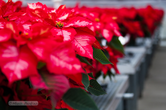
A small portion of one of five greenhouses in Orange Coast College's Horticulture Department that are filled with poinsettias being grown for their annual 2011 poinsettia sale.  This image focuses on a single ice punch poinsettia, highlighting how the red and white leaves are nothing more than bracts - colored leaves grown near a flower.  The actual flowers are the green, red, and yellow structures at the top of the plant (end of the stem). (Marc C. Perkins)
A small portion of one of five greenhouses in Orange Coast College's Horticulture Department that are filled with poinsettias being grown for their annual 2011 poinsettia sale. The plants in the foreground are the "Ice Punch" variety.

Many people think that the big red (or red and white, in the picture above) structures are the flower petals of poinsettias; they’re not.  The big red structures are bracts: specialized leaves associated with a flower that are typically brightly colored (usually to help attract pollinators to inconspicuous flowers).  The actual flowers of the plant are located at the ends of the stems, and are significantly smaller and less noticeable.  Look closely at the picture above, though, and you’ll see little green, red, and yellow clusters at the top of the plant; those are the flowers.

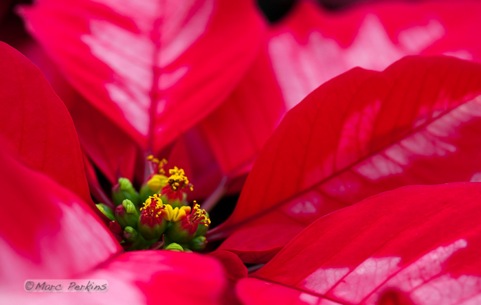
A closeup of a poinsettia flower cluster from the side. Many people confuse the bracts (red and white leaves on this "Ice Punch" poinsettia) with the petals of flowers; they're quite different.   This macro shot shows a number of bracts (colored leaves associated with a flower) surrounding the flowers (green, red, and yellow structures) and extending out of the frame.  The flowers themselves are called cyanthia; the green tissue surrounding each flower is an involucre, a cluster of bracts (leaves) fused into a cup-shaped structure that contains multiple male flowers and one female flower within it.  Emerging from the involucre you can see red filaments supporting yellow anthers on the male flowers; a single female flower should be emerging from the center of each flower.  The bright yellow structures attached to the involucre are nectar glands filled with yummy sweet nectar to attract pollinators; on less-developed flowers they look like little light-green lips. (Marc C. Perkins)
A closeup of an Ice Punch poinsettia flower cluster from the side.

In the picture above we can see that the true flowers of the poinsettia are not the brightly colored leaves, but instead are these small green and red structures. The yellow things that look like lips are nectar glands, and the green balls with red filaments are the flowers (which are more appropriately termed pseudanthia or inflorescences, since they’re actually multiple flowers in a single structure). Let’s look at them closer:

A closeup of a poinsettia flower cluster from directly above it. Many people confuse the bracts (red leaves) with the petals of flowers; they're quite different.   This macro shot shows a number of bracts (colored leaves associated with a flower) surrounding the flowers (green, red, and yellow structures) and extending out of the frame.  The inflorescences are called cyanthia; the green tissue surrounding each one is an involucre, a cluster of bracts (leaves) fused into a cup-shaped structure that contains multiple male flowers and one female flower within it.  Emerging from the involucre you can see red filaments supporting yellow anthers on the male flowers; a single female flower should be emerging from the center of each flower.  The bright yellow, liquid-filled structures attached to the involucre are nectar glands filled with yummy sweet nectar to attract pollinators; on less-developed flowers they look like little light-green lips. (Marc C. Perkins)
A closeup of a poinsettia flower cluster from directly above. Many people confuse the bracts (red leaves) with the petals of flowers; they're quite different.

This macro shot shows a number of red bracts surrounding the flowers and extending out of the frame. The green balls tipped with red are individual inflorescences called cyanthia, which are composed of multiple flowers surrounded by modified leaves1. The green tissue surrounding each inflorescence is an involucre, a cluster of bracts (modified leaves) fused into a cup-shaped structure that contains multiple male flowers and one female flower within it.

Emerging from each involucre are red filaments supporting yellow anthers that are being grown by the male flowers (the anthers produce and release pollen).  A single female flower should be emerging from the center of each involucre, but isn’t easily visible in the picture.

The yellow liquid-filled structures attached to each involucre are nectar glands filled with nectar to attract pollinators.  On less-developed inflorescences the nectar glands look like little light-green lips.

If you’re in the Orange County area and want your own poinsettias to observe the flowers of, head to OCC this Friday and pick some up for yourself!  You’ll get a cool botanical specimen, and will be supporting a great program in the process. Oh yeah, and you’ll have a nice pretty plant for the holiday season.

[This is the first in a series of three posts; the second post is: Poinsettia flower closeups take two: getting even closer and the third is Poinsettia flowers part three: the female parts.]

1 Yes, the plant works hard to hide its flowers. First there are dozens of large colorful bracts, and then smaller green ones. Sheesh!

Spiders in the night

It’s fall here in coastal Orange County, CA1, but determining that it’s fall can be difficult since we don’t have trees filled with yellow and red leaves.  Around here I find that there’s no better indicator of fall than seeing one of these hanging around outside2:

A large orb weaver spider (genus _Araneus_) sits in the center of her web, just waiting for some prey (you, perhaps?) to come along and get stuck in her web.  All eight legs, her giant abdomen, and her pedipalps are nicely visible, as is the central portion of her web.  These spiders spin their webs at dusk, hence the nighttime black background.  I love how her legs are spread out so that they connect to more than half of the major supporting strands of the web.  And yes, she was facing down, towards the ground. (Marc C. Perkins)
A large orb weaver spider (genus _Araneus_) sits in the center of her web, just waiting for some prey (you, perhaps?) to come along and get stuck in her web.

These orb weaver spiders come out every year in late summer and early fall, building webs at dusk that are frequently more than a foot across and can have individual lines of silk running more than 10 feet from attachment point to attachment point.  They’re amazing animals, and I love to see them every year3.

This year a few took up residence close enough for me to try out my new macro lens.  And, since it’s almost Halloween, it’s a perfect time to post up some spider pictures.  Here’s one of the spiders just hangin’ out:

An orb weaver spider (genus _Araneus_) hangs upside down from a portion of its web.  The large and hairy black and white abdomen is clearly visible. (Marc C. Perkins)
An orb weaver spider (genus _Araneus_) hangs upside down from a portion of its web.

In this profile shot you can see how the spider has a small strand of silk attaching herself to the web as a safety-strap:

A large tan orb weaver spider (genus _Araneus_) hangs down from her web in this side-view (profile) of her.  She's hanging from intact strands of her web, but the strands above those (that are all coiled up) are from a portion of the web that was damaged.  I like how you can see a single strand of spider silk running from her spinneretes to the web, acting as a safety strap. (Marc C. Perkins)
A large tan orb weaver spider (genus _Araneus_) hangs down from her web in this side-view (profile) of her. She's hanging from intact strands of her web, but the strands above those (that are all coiled up) are from a portion of the web that was damaged.

The attachment points of all eight legs onto the bottom of the cephalothorax is a fun feature to focus on (and probably the last view of many a doomed insect):

Continue reading Spiders in the night

Photographing mice: the adorableness is overwhelming!

A gray male pet mouse stands on all four paws and looks just past the camera, seemingly ready for anything.  He's in an exploration mode, looking around the area with ears perked up and whiskers at the ready. (Marc C. Perkins)
A gray male pet mouse stands on all four paws and looks just past the camera, seemingly ready for anything.

Recently I got the opportunity to photograph a few fancy mice, and I just have to share the cuteness here on the blog. I had rats or mice as pets for more than a decade before getting my current cats, and I dearly loved them; in fact, I’d still have rodent pets if it weren’t for their horribly short lifespans 1.

Editor’s note: I’ll try to add commentary throughout these pictures, but I should probably just say “SQUEEE!” after each picture, because that’s what I’m really doing inside when I see these.

A gray male mouse with black eyes peers over the top of a clear plastic cage.  He's just poking his nose over, and looks like he's hesitantly wondering what, or who, is out there. (Marc C. Perkins)
A gray male mouse with black eyes peers over the top of a clear plastic cage. He's just poking his nose over, and looks like he's hesitantly wondering what, or who, is out there.

Mice are incredibly fun to watch, especially in large groups. They’re always exploring the boundaries of their cages:

A gray male pet mouse jumps up on the side of a clear plastic cage and holds himself up by his front paws, peering over the edge into the vast unknown beyond the cage.  I love the cute little paws holding onto the edge.  I also like how the whiskers are fully three-dimensional: you can see how they extend around the face in all directions - front, back, top, bottom, and sides. (Marc C. Perkins)
A gray male pet mouse jumps up on the side of a clear plastic cage and holds himself up by his front paws, peering over the edge into the vast unknown beyond the cage. I like how the whiskers are fully three-dimensional: you can see how they extend around the face in all directions - front, back, top, bottom, and sides.

or poking their noses out of tubes:

A white and black patched male mouse sniffs at what's outside his green tube.  The focus is dead on his adorable pink nose! (Marc C. Perkins)
A white and black patchy male mouse sniffs at what's outside his green tube. The focus is right on his adorable pink nose!

or grooming themselves:

Continue reading Photographing mice: the adorableness is overwhelming!

The insects and spiders of Crystal Cove State Park: bees, butterflies, and more!

I’ve already posted a few pictures from my recent backpacking trip to Crystal Cove State Park (flowers, self-portrait, rattlesnake mating). But as I was photographing dew-covered flowers on the foggy morning, I couldn’t help but notice the spiders in their spiderwebs.

This orb weaving spider is standing so that it appears to have only four legs.  Its cephalothorax is also covered in fine white hairs.  Seen on a beautifully foggy morning in Crystal Cove State Park, there are a few drops of dew on the spider itself and its web is covered in drops.   My best guess is that this is in genus _Argiope_.  I have a shot of this with better depth of field (all of the web's water droplets are in focus), if you would prefer that (but the background becomes distracting). (Marc C. Perkins)
An orb weaving spider in a dew covered web standing so that it appears to have only four legs. My best guess is that this is in genus _Argiope_.

I had to get up closer, of course:

This orb weaving spider is standing so that it appears to have only four legs.  Is it trying to pretend to be a vertebrate? Its cephalothorax is also covered in fine white hairs.  Seen on a beautifully foggy morning in Crystal Cove State Park, there are a few drops of dew on the spider itself.   My best guess is that this is in genus _Argiope_. (Marc C. Perkins)
Up close and personal, this spider (probably in _Argiope_) is just gorgeous on a foggy morning.

And, you know me, I’m not satisfied unless the subject is spilling out of the frame, but I’ll spare you from a third picture of the same spider in the same post.

Since I know spiders are everyone’s favorite critter, here’s another one I saw that morning:

A brown and tan orb weaver spider stands on its dew covered web in front of an orange background. (Marc C. Perkins)
A brown and tan orb weaver spider stands on its dew covered web in front of an orange background.

But I saw more than just spiders that morning. After the fog burned off, the day warmed up and a ton of insects came out to go about their business. Chief among them were dozens of bees buzzing around this plant:

A honeybee (_Apis mellifera_) forager samples the wares at an all-you-can-eat buffet of white and purple/yellow spike-inflorescence flowers in Crystal Cove State Park. (Marc C. Perkins)
A honeybee (_Apis mellifera_) forager samples the wares at an all-you-can-eat buffet of white and purple/yellow spike-inflorescence flowers in Crystal Cove State Park.

While most of the spider and flower shots I’ve already posted used my new macro lens on a tripod, the rest of theses shots tested out how the lens performed hand-held with fast-moving subjects. It worked very well, auto-focusing quickly and allowing me to get decent depth of field with shutter speeds fast enough to freeze motion:

A honeybee (_Apis mellifera_) forager sticks her antennae and forelegs into a small purple and white flower at Crystal Cove State Park.  The bee's pollen basket (corbicula) on her hind legs are stuffed full with tan pollen. (Marc C. Perkins)
A honeybee (_Apis mellifera_) forager sticks her antennae and forelegs into a small purple and white flower at Crystal Cove State Park. The bee's pollen basket (corbicula) on her hind legs is stuffed full with tan pollen.

Of course there were more than just bees and spiders out. My hiking partners spotted this cabbage butterfly flitting around on the trail, and the macro exceeded my expectations by allowing me to capture this image in the few seconds I had before the butterfly flitted away 1:

Continue reading The insects and spiders of Crystal Cove State Park: bees, butterflies, and more!

Location alert: Flowering agaves at Little Corona Beach

This past Monday I found myself in possession of a rare bit of spare time around sunset, so I zipped down to Little Corona Beach, which is just off Pacific Coast Highway in Corona Del Mar. My primary purpose for the trip was to try out my new three-stop neutral density filter, but I found a nice surprise at the entrance to the beach:

Two flowering agaves on the bluffs at Little Corona Beach, seen just before sunset.  I love the combination of classic California coastal bluffs in the background and the inflorescences in the foreground.  However, I don't like the sign in the foreground ... (Marc C. Perkins)
Two flowering agaves on the bluffs at Little Corona Beach, seen just before sunset. I love the combination of classic California coastal bluffs in the background and the inflorescences in the foreground.

The agaves were absolutely gorgeous, especially since their quickly-growing inflorescences could be composed with classic California ocean scenes in the background.

Two flowering agaves on the bluffs at Little Corona Beach, seen just before sunset.  I love the combination of classic California coastal bluffs in the background and the inflorescences in the foreground. (Marc C. Perkins)
Two flowering agaves on the bluffs at Little Corona Beach, seen just before sunset.

These inflorescences won’t be around forever. Just like the Agave I photographed earlier this year (gallery 1, gallery 2), the inflorescence will grow to a certain height, burst into flower, and then die (and then likely get ripped out by the landscapers, which is what happened to the one at OCC). Also, agaves typically flower only once during their entire lives.

So, local photographers, this is your only chance to capture these agaves at their peak. I’m no botanist, but my guess is that they’ll quickly grow too tall for their tips to be included in a sea-scape composition without a ladder. I don’t know how long it will be until they flower, either, but my guess is we’ve got a few weeks (as I didn’t see any flower buds present yet).

If you do go and photograph them, I’ve got only two requests:

  1. Respect the fence that protects the agaves and their surrounding vegetation. The bluffs are very delicate, and local groups have been working very hard to re-vegetate the area.
  2. If you do get some good shots, I’d love it if you could share them with everyone by posting a comment here linking to them.

Continue reading Location alert: Flowering agaves at Little Corona Beach

Rattlesnake mating: frantic sensuality in the grass

[Update February 28, 2012: Thanks to a most-excellent comment by Bryan Hughes, I now know that this is a post about aggression between two male rattlesnakes, not a male and female snake mating.]

A few weeks ago some friends and I camped overnight in Crystal Cove’s Lower Moro Campground. The morning was beautifully foggy, and I took a bunch of flower closeups while the dew was still around. But the sun eventually burned through the fog, and we packed up and headed for home.

As I was hiking I heard some rustling in the grass next to me beside the trail. Accustomed to hunting for lizards, I turned and looked to see what it was. Lizard? Bird? Rodent?

Rattlesnake.

No, wait.

Two rattlesnakes.

About that time my legs decided that they really didn’t like being about two feet from two writhing rattlesnakes, so my hiking partners and I quickly moved away. But luckily for us, the snakes were in the throes of passion, and didn’t seem to notice us at all. So, I pulled out my camera and started snapping.

My favorite shot of the bunch is this one:

I'm pretty sure these are two southern pacific rattlesnakes (_Crotalus oreganus_ helleri) mating.  I first noticed the pair when I was only a foot or two away from them on a trail; after moving a bit away I captured this shot of the two snakes wrapped around each other.  Sadly, I think my presence may have interrupted them. (Marc C. Perkins)
Two southern pacific rattlesnakes (_Crotalus oreganus_ helleri) mating. Click on the image to see it larger.

There the two have just picked themselves up after reaching up vertically and coiling around each other so much that they fell over. To see the entire sequence of them coiling, here’s a five-image composite:

A five image sequence of two southern pacific rattlesnakes (_Crotalus oreganus_ helleri) mating.  I first noticed the pair when I was only a foot or two away from them on a trail; after moving a bit away I captured these five shots of the two snakes wrapping around each other.  The top four images were captured over no more than 2 seconds total, and the large image was taken less than 7 seconds after the top four. (Marc C. Perkins)
A five image sequence of two southern pacific rattlesnakes (_Crotalus oreganus_ helleri) mating. The top four images were captured over no more than 2 seconds total, and the large image was taken less than 7 seconds after the top four. Click on the image to see it larger.

Shortly after that, however, either they were done or they noticed us. One of them slithered off into the bushes, while the other almost seemed to stand guard, staring out at us with tongue flicking.

Shortly after the amazingly-fast coiling seen in my prior images, the two snakes separated.  One slithered off into the brush, while the other started paying just a bit too much attention to me and my colleagues for my tastes.  So, we decided to part ways amicably; I even got a nice rattle as the snake slithered away. (Marc C. Perkins)
Shortly after the amazingly fast coiling seen in my prior images, the two snakes separated. One slithered off into the brush, while the other started paying just a bit too much attention to me and my colleagues for my taste. Click on the image to see it larger.

We took the hint and started to move off, and the guard snake did the same, giving us a nice rattle as it crawled into the brush.

It was easily one of the most exciting animal encounters I’ve had while hiking or backpacking. They were incredibly fast as they coiled and writhed, and their ritual was entrancingly beautiful.

Continue reading Rattlesnake mating: frantic sensuality in the grass

Flowers of Crystal Cove State Park: The joys of a foggy morning

A few weeks ago I went backpacking with a couple of friends in Crystal Cove State Park’s inland section. We camped overnight at the Lower Moro Campground, and were the only ones there. It was wonderfully peaceful.

Fog rolled in a few hours before the sun went down, so sunset, moon, and starry night shots were all out. But the fog hung around until well after dawn, leaving everything covered in a beautiful shroud of dew the next morning. It made for perfect macro photography, and my companions were patient enough to let me spend some time trying to capture the beauty.

I woke up to a foggy morning at Lower Moro campground in Crystal Cove, and found this beautiful little purple and white flower covered in dew.  I love the few strands of spider silk connecting the flower to its stalk, also covered in water droplets.  A botanist friend of mine identified this as _Stephanomeria sp._, a plant in Asteraceae. (Thanks Jeremy!) (Marc C. Perkins)
A beautiful way to wake up. I love the few strands of dew-covered spider silk connecting the flower to its stalk. A botanist friend of mine identified this as wreath plant, _Stephanomeria sp._, a plant in Asteraceae. (Thanks Jeremy!)

The same plant was also covered in tiny flower buds, which the dew accentuated gorgeously:

This small green and purple flower bud growing out of a stem is covered in dew on a foggy morning at Crystal Cove, and the dew is lensing the background into focus.  The few strands of spider silk are also covered in dew. A botanist friend of mine identified this as wreath plant, _Stephanomeria sp._, a plant in Asteraceae. (Thanks Jeremy!)
A small green and purple flower bud (_Stephanomeria sp._) covered in dew on a foggy morning at Crystal Cove.

I also got a picture of the same plant with both a flower bud and flower in the same frame, but I won’t bore you with that in this post.

Nearby there was a shrubby aster covered in bright yellow flowers:

This yellow Asteraceae flower is a composite flower (capitulum)consisting of multiple individual flowers attached to the same base.  Here the flowers on the outside of the composite flower (the ray florets) are open, but the inner flowers (the disk florets) are still closed.  The ray florets fuse their petals into one giant lobe.  See the next picture to see the same plant's flower with the disk florets open. This is most likely one of the several species of tarplant, possibly _Deinandra_. (Marc C. Perkins)
This yellow Asteraceae flower is a composite flower (capitulum) consisting of multiple individual flowers attached to the same base. Here the flowers on the outside of the composite flower (the ray florets) are open, but the inner flowers (the disk florets) are still closed. The ray florets fuse their petals into one giant lobe. This is most likely one of the several species of tarplant, possibly _Deinandra_.

Some of the inflorescences were just starting to open (above), while others were almost fully open (below):

Continue reading Flowers of Crystal Cove State Park: The joys of a foggy morning