Tag Archives: macro

Poinsettia flower closeups take two: getting even closer

[This is the second in a series of three posts; the first post is: Poinsettias up close: where are the flowers? and the third is Poinsettia flowers part three: the female parts.]

A closeup of a poinsettia flower cluster from directly above it. Many people confuse the bracts (red leaves) with the petals of flowers; they're quite different.   This macro shot shows a number of bracts (colored leaves associated with a flower) surrounding the flowers (green, red, and yellow structures) and extending out of the frame.  The flowers themselves are called cyanthia; the green tissue surrounding each flower is an involucre, a cluster of bracts (leaves) fused into a cup-shaped structure that contains multiple male flowers and one female flower within it.  Emerging from the involucre you can see red filaments supporting yellow anthers on the male flowers; a single female flower should be emerging from the center of each flower.  The bright yellow, liquid-filled structures attached to the involucre are nectar glands filled with yummy sweet nectar to attract pollinators; on less-developed flowers they look like little light-green lips. (Marc C. Perkins)
A closeup of a poinsettia flower cluster from directly above. This macro image shows a number of inflorescences (green, red, and yellow structures) surrounded by bracts (colored leaves associated with a flower) extending out of the frame.

Earlier this week I posted about finding the flowers in poinsettias (spoiler: the petals aren’t the big red structures!).  After writing that post, though, I realized it was missing a good closeup of the flower clusters themselves.  So, yesterday afternoon I took a quick break from my end-of-the-semester piles of grading and headed back to Orange Coast College’s Ornamental Horticulture Department to get a few more pictures.

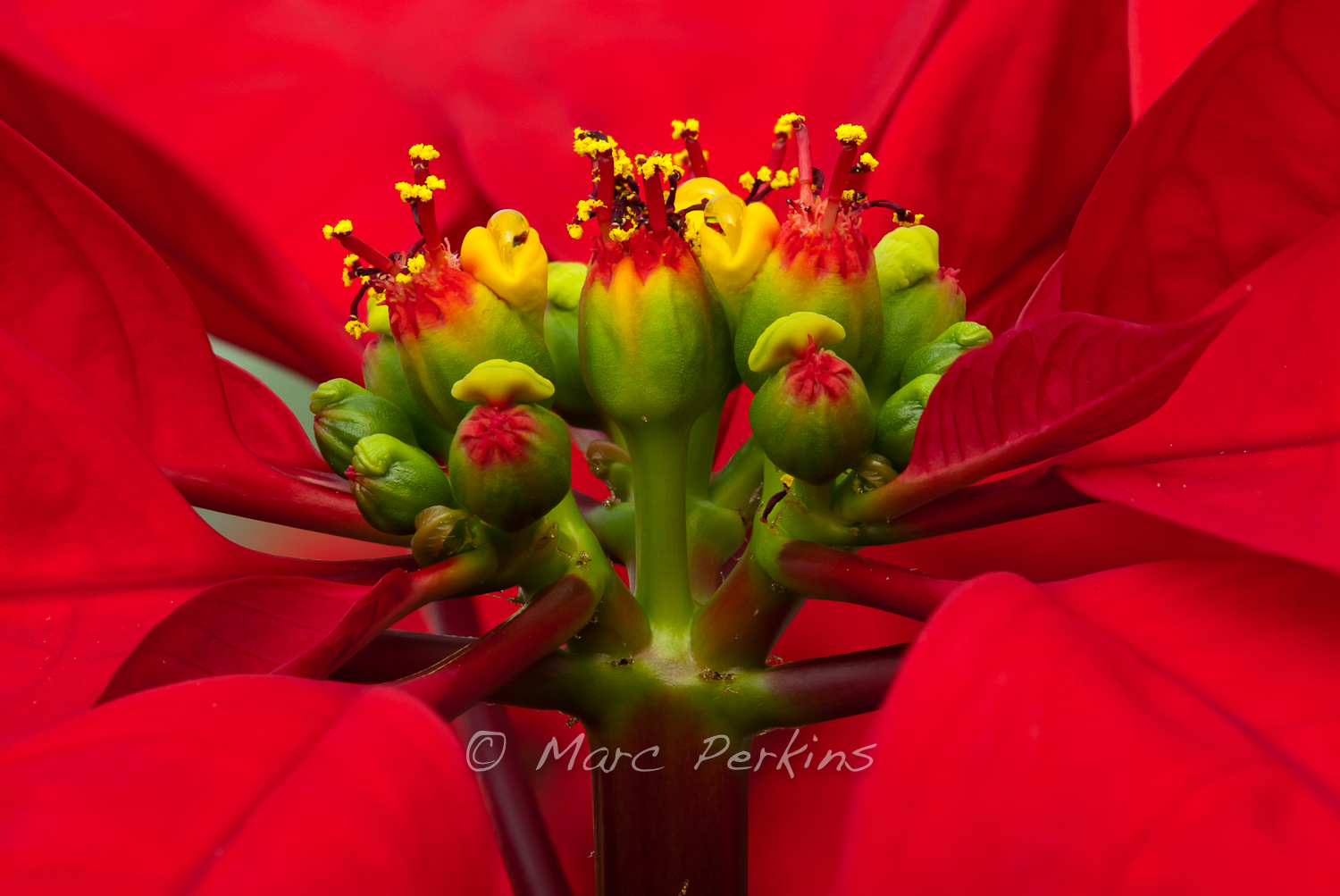
The flowers of poinsettias (Euphorbia pulcherrima) are actually cyanthia: flowering structures composed of many individual male or female flowers surrounded by modified leaves.  Here’s what they look like:

A closeup of a poinsettia flower cluster from the side.  Many people confuse the bracts (red leaves) with the actual flowers; they're quite different.   This macro shot shows multiple pseudanthia surrounded by a sea of red bracts (colored leaves associated with a flower).  The flowers themselves are called cyanthia; the green tissue surrounding each flower is an involucre, a cluster of bracts (leaves) fused into a cup-shaped structure that contains multiple male flowers and one female flower within it.  Emerging from the involucre you can see red filaments supporting yellow anthers on the male flowers, and you can even see some of the pollen grains.  Also emerging from each involucre you can see a number of dark-purple structures supported by short stalks (that are about a tenth of the height of the filaments; I'm not sure what these are).   The bright yellow, liquid-filled structures attached to the involucre are nectar glands filled with nectar to attract pollinators. (Marc C. Perkins)
A closeup of a poinsettia flower cluster from the side, showing how the cyanthia branch off of the tip of a stem and are surrounded by red bracts.

To see that image larger, follow this link to see it as a large, high-resolution image.

That picture shows multiple cyanthia (flower clusters) at the end of a stem surrounded by a sea of red bracts (colored leaves associated with a flower). Each green ball tipped with red is an involucre, a cluster of bracts (leaves) fused into a cup-shaped structure that contains multiple male flowers and one female flower within it.

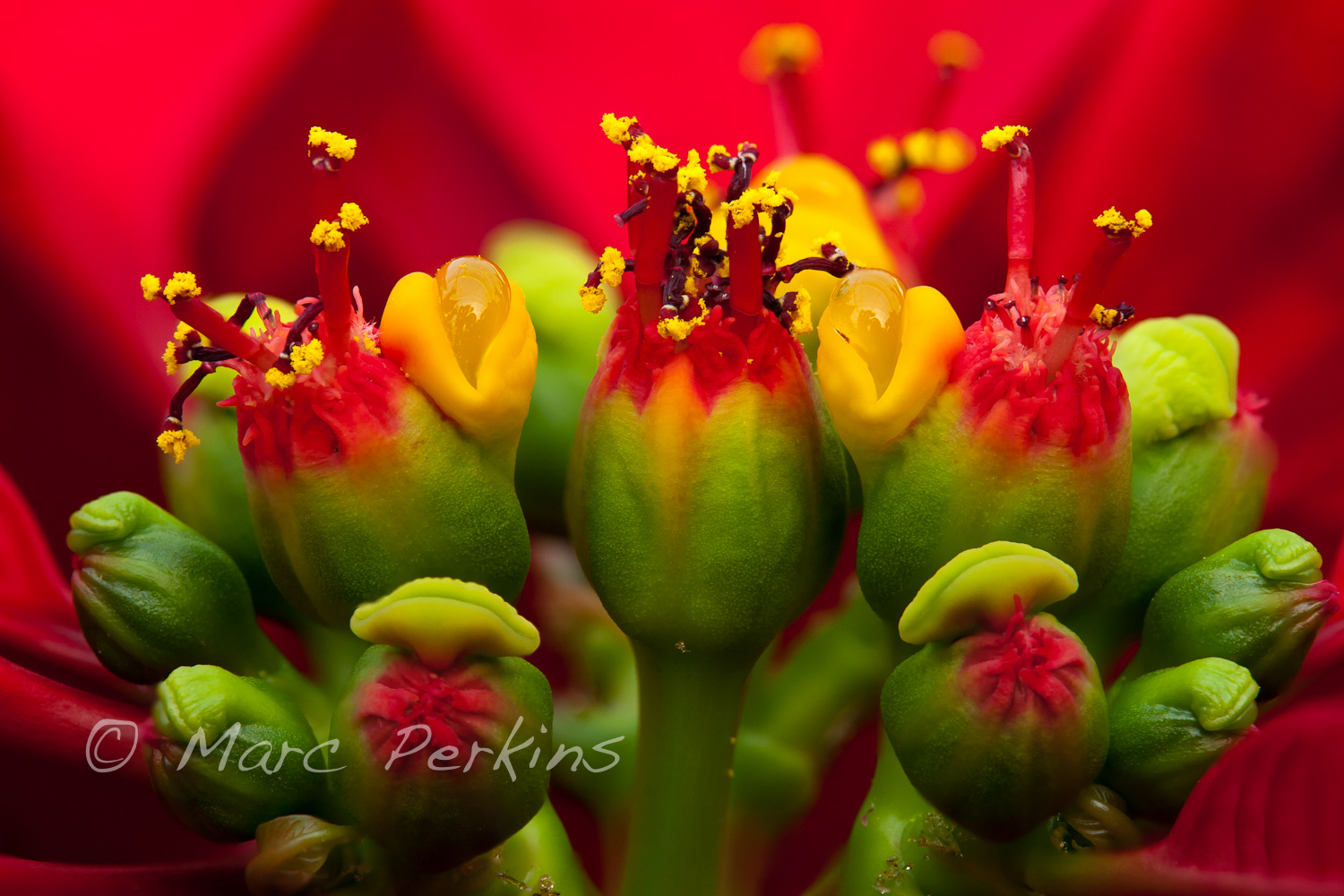
Let’s look at the involucres and their flowers even closer:

A closeup of a poinsettia flower cluster from the side.  Many people confuse the bracts (red leaves) with the actual flowers; they're quite different.   This macro shot shows multiple pseudanthia surrounded by a sea of red bracts (colored leaves associated with a flower; mostly out of focus in this picture).  The flowers themselves are called cyanthia; the green tissue surrounding each flower is an involucre, a cluster of bracts (leaves) fused into a cup-shaped structure that contains multiple male flowers and supposedly one female flower within it, though I see multiple stigma in each involucre.  Emerging from the involucre you can see red filaments supporting yellow anthers on the male flowers, and you can even see some of the pollen grains.  Also emerging from each involucre you can see a number of dark-purple stigmas supported by short styles (that are about a tenth of the height of the filaments).   The bright yellow, liquid-filled structures attached to the involucre are nectar glands filled with nectar to attract pollinators. (Marc C. Perkins)
A closeup of a poinsettia flower cluster from the side. Follow the link below to see the image at twice the size.

To see that image in all its glory, follow this link to see it as a large, high-resolution image.

Emerging from each involucre you can see red filaments supporting yellow anthers that are dusty with individual pollen grains.  The filaments are emerging from the multiple male flowers growing within each involucre.   Also emerging from each involucre you can see a number of dark-purple structures supported by short stalks; I believe these may be the stigmas and styles of the flowers (though this species is supposed to have only a single female flower per involucre with a stigma divided into three sections, so I’m not certain what those dark-purple structures are).

[edited 1/20/2012: the dark purple structures are indeed not the female flowers, as I write about in this post. I’m not at all sure what these small structures are. Anyone have any ideas?]

The bright yellow, liquid-filled structures attached to the involucre are nectar glands filled with nectar (to attract pollinators).  A few individual pollen grains are stuck to the surface of the left-most nectar gland.

I’ll leave you with a crop of that last image showing two cyanthia in more detail

A sample of the detail visibile in my "Poinsettia inflorescence extreme closeup".  To see the full image or purchase a copy, go to that image.  That picture is a closeup of a poinsettia flower cluster from the side.  Many people confuse the bracts (red leaves) with the actual flowers; they're quite different.   This macro shot shows multiple pseudanthia surrounded by a sea of red bracts (colored leaves associated with a flower; mostly out of focus in this picture).  The flowers themselves are called cyanthia; the green tissue surrounding each flower is an involucre, a cluster of bracts (leaves) fused into a cup-shaped structure that contains multiple male flowers and supposedly one female flower within it, though I see multiple stigma in each involucre.  Emerging from the involucre you can see red filaments supporting yellow anthers on the male flowers, and you can even see some of the pollen grains.  Also emerging from each involucre you can see a number of dark-purple stigmas supported by short styles (that are about a tenth of the height of the filaments).   The bright yellow, liquid-filled structures attached to the involucre are nectar glands filled with nectar to attract pollinators. (Marc C. Perkins)
Two cyanthia of a poinsettia flower cluster, with anthers and associated structures easily visible.  This is a sample of the detail visibile in the image posted above.

And, if you want your own poinsettias to admire, head to Orange Coast College this Friday: they’re having their annual poinsettia sale, which is open to the public.  See my first post in this series for more information.

More pictures

To see more of my poinsettia pictures, head to my Poinsettia Gallery.

Poinsettias up close: where are the flowers?

[This is the first in a series of three posts; the second post is: Poinsettia flower closeups take two: getting even closer and the third is Poinsettia flowers part three: the female parts.]

Orange Coast College’s Ornamental Horticulture Department is just plain awesome. The department’s landscaped gardens are easily the most beautiful spot on campus, they teach a wide array of neat classes (landscape design, plant propagation, cacti and succulents, etc.), and they’ve got the nicest faculty and staff around.

To help fund their department, and provide a lab opportunity for their plant propagation class, every fall the department rears thousands of poinsettias (Euphorbia pulcherrima) in their greenhouses for sale to the public. This year’s sale is this Friday (December 9), and their greenhouses are currently full of beautiful poinsettias.

I just had to take a look at the plants last week:

A small portion of one of five greenhouses in Orange Coast College's Horticulture Department that are filled with poinsettias being grown for their annual 2011 poinsettia sale.  This image focuses on a single ice punch poinsettia, highlighting how the red and white leaves are nothing more than bracts - colored leaves grown near a flower.  The actual flowers are the green, red, and yellow structures at the top of the plant (end of the stem). (Marc C. Perkins)
A small portion of one of five greenhouses in Orange Coast College's Horticulture Department that are filled with poinsettias being grown for their annual 2011 poinsettia sale. The plants in the foreground are the "Ice Punch" variety.

Many people think that the big red (or red and white, in the picture above) structures are the flower petals of poinsettias; they’re not.  The big red structures are bracts: specialized leaves associated with a flower that are typically brightly colored (usually to help attract pollinators to inconspicuous flowers).  The actual flowers of the plant are located at the ends of the stems, and are significantly smaller and less noticeable.  Look closely at the picture above, though, and you’ll see little green, red, and yellow clusters at the top of the plant; those are the flowers.

A closeup of a poinsettia flower cluster from the side. Many people confuse the bracts (red and white leaves on this "Ice Punch" poinsettia) with the petals of flowers; they're quite different.   This macro shot shows a number of bracts (colored leaves associated with a flower) surrounding the flowers (green, red, and yellow structures) and extending out of the frame.  The flowers themselves are called cyanthia; the green tissue surrounding each flower is an involucre, a cluster of bracts (leaves) fused into a cup-shaped structure that contains multiple male flowers and one female flower within it.  Emerging from the involucre you can see red filaments supporting yellow anthers on the male flowers; a single female flower should be emerging from the center of each flower.  The bright yellow structures attached to the involucre are nectar glands filled with yummy sweet nectar to attract pollinators; on less-developed flowers they look like little light-green lips. (Marc C. Perkins)
A closeup of an Ice Punch poinsettia flower cluster from the side.

In the picture above we can see that the true flowers of the poinsettia are not the brightly colored leaves, but instead are these small green and red structures. The yellow things that look like lips are nectar glands, and the green balls with red filaments are the flowers (which are more appropriately termed pseudanthia or inflorescences, since they’re actually multiple flowers in a single structure). Let’s look at them closer:

A closeup of a poinsettia flower cluster from directly above it. Many people confuse the bracts (red leaves) with the petals of flowers; they're quite different.   This macro shot shows a number of bracts (colored leaves associated with a flower) surrounding the flowers (green, red, and yellow structures) and extending out of the frame.  The inflorescences are called cyanthia; the green tissue surrounding each one is an involucre, a cluster of bracts (leaves) fused into a cup-shaped structure that contains multiple male flowers and one female flower within it.  Emerging from the involucre you can see red filaments supporting yellow anthers on the male flowers; a single female flower should be emerging from the center of each flower.  The bright yellow, liquid-filled structures attached to the involucre are nectar glands filled with yummy sweet nectar to attract pollinators; on less-developed flowers they look like little light-green lips. (Marc C. Perkins)
A closeup of a poinsettia flower cluster from directly above. Many people confuse the bracts (red leaves) with the petals of flowers; they're quite different.

This macro shot shows a number of red bracts surrounding the flowers and extending out of the frame. The green balls tipped with red are individual inflorescences called cyanthia, which are composed of multiple flowers surrounded by modified leaves1. The green tissue surrounding each inflorescence is an involucre, a cluster of bracts (modified leaves) fused into a cup-shaped structure that contains multiple male flowers and one female flower within it.

Emerging from each involucre are red filaments supporting yellow anthers that are being grown by the male flowers (the anthers produce and release pollen).  A single female flower should be emerging from the center of each involucre, but isn’t easily visible in the picture.

The yellow liquid-filled structures attached to each involucre are nectar glands filled with nectar to attract pollinators.  On less-developed inflorescences the nectar glands look like little light-green lips.

If you’re in the Orange County area and want your own poinsettias to observe the flowers of, head to OCC this Friday and pick some up for yourself!  You’ll get a cool botanical specimen, and will be supporting a great program in the process. Oh yeah, and you’ll have a nice pretty plant for the holiday season.

[This is the first in a series of three posts; the second post is: Poinsettia flower closeups take two: getting even closer and the third is Poinsettia flowers part three: the female parts.]

1 Yes, the plant works hard to hide its flowers. First there are dozens of large colorful bracts, and then smaller green ones. Sheesh!

Spiders in the night

It’s fall here in coastal Orange County, CA1, but determining that it’s fall can be difficult since we don’t have trees filled with yellow and red leaves.  Around here I find that there’s no better indicator of fall than seeing one of these hanging around outside2:

A large orb weaver spider (genus _Araneus_) sits in the center of her web, just waiting for some prey (you, perhaps?) to come along and get stuck in her web.  All eight legs, her giant abdomen, and her pedipalps are nicely visible, as is the central portion of her web.  These spiders spin their webs at dusk, hence the nighttime black background.  I love how her legs are spread out so that they connect to more than half of the major supporting strands of the web.  And yes, she was facing down, towards the ground. (Marc C. Perkins)
A large orb weaver spider (genus _Araneus_) sits in the center of her web, just waiting for some prey (you, perhaps?) to come along and get stuck in her web.

These orb weaver spiders come out every year in late summer and early fall, building webs at dusk that are frequently more than a foot across and can have individual lines of silk running more than 10 feet from attachment point to attachment point.  They’re amazing animals, and I love to see them every year3.

This year a few took up residence close enough for me to try out my new macro lens.  And, since it’s almost Halloween, it’s a perfect time to post up some spider pictures.  Here’s one of the spiders just hangin’ out:

An orb weaver spider (genus _Araneus_) hangs upside down from a portion of its web.  The large and hairy black and white abdomen is clearly visible. (Marc C. Perkins)
An orb weaver spider (genus _Araneus_) hangs upside down from a portion of its web.

In this profile shot you can see how the spider has a small strand of silk attaching herself to the web as a safety-strap:

A large tan orb weaver spider (genus _Araneus_) hangs down from her web in this side-view (profile) of her.  She's hanging from intact strands of her web, but the strands above those (that are all coiled up) are from a portion of the web that was damaged.  I like how you can see a single strand of spider silk running from her spinneretes to the web, acting as a safety strap. (Marc C. Perkins)
A large tan orb weaver spider (genus _Araneus_) hangs down from her web in this side-view (profile) of her. She's hanging from intact strands of her web, but the strands above those (that are all coiled up) are from a portion of the web that was damaged.

The attachment points of all eight legs onto the bottom of the cephalothorax is a fun feature to focus on (and probably the last view of many a doomed insect):

Continue reading Spiders in the night

Flowers of Crystal Cove State Park: The joys of a foggy morning

A few weeks ago I went backpacking with a couple of friends in Crystal Cove State Park’s inland section. We camped overnight at the Lower Moro Campground, and were the only ones there. It was wonderfully peaceful.

Fog rolled in a few hours before the sun went down, so sunset, moon, and starry night shots were all out. But the fog hung around until well after dawn, leaving everything covered in a beautiful shroud of dew the next morning. It made for perfect macro photography, and my companions were patient enough to let me spend some time trying to capture the beauty.

I woke up to a foggy morning at Lower Moro campground in Crystal Cove, and found this beautiful little purple and white flower covered in dew.  I love the few strands of spider silk connecting the flower to its stalk, also covered in water droplets.  A botanist friend of mine identified this as _Stephanomeria sp._, a plant in Asteraceae. (Thanks Jeremy!) (Marc C. Perkins)
A beautiful way to wake up. I love the few strands of dew-covered spider silk connecting the flower to its stalk. A botanist friend of mine identified this as wreath plant, _Stephanomeria sp._, a plant in Asteraceae. (Thanks Jeremy!)

The same plant was also covered in tiny flower buds, which the dew accentuated gorgeously:

This small green and purple flower bud growing out of a stem is covered in dew on a foggy morning at Crystal Cove, and the dew is lensing the background into focus.  The few strands of spider silk are also covered in dew. A botanist friend of mine identified this as wreath plant, _Stephanomeria sp._, a plant in Asteraceae. (Thanks Jeremy!)
A small green and purple flower bud (_Stephanomeria sp._) covered in dew on a foggy morning at Crystal Cove.

I also got a picture of the same plant with both a flower bud and flower in the same frame, but I won’t bore you with that in this post.

Nearby there was a shrubby aster covered in bright yellow flowers:

This yellow Asteraceae flower is a composite flower (capitulum)consisting of multiple individual flowers attached to the same base.  Here the flowers on the outside of the composite flower (the ray florets) are open, but the inner flowers (the disk florets) are still closed.  The ray florets fuse their petals into one giant lobe.  See the next picture to see the same plant's flower with the disk florets open. This is most likely one of the several species of tarplant, possibly _Deinandra_. (Marc C. Perkins)
This yellow Asteraceae flower is a composite flower (capitulum) consisting of multiple individual flowers attached to the same base. Here the flowers on the outside of the composite flower (the ray florets) are open, but the inner flowers (the disk florets) are still closed. The ray florets fuse their petals into one giant lobe. This is most likely one of the several species of tarplant, possibly _Deinandra_.

Some of the inflorescences were just starting to open (above), while others were almost fully open (below):

Continue reading Flowers of Crystal Cove State Park: The joys of a foggy morning

Photographing lichen: using a hand-held reflector to get better lighting

Lichen are visually fascinating: their bodies have gorgeous textures, lines, and colors. But capturing all of that can be challenging. Take, for instance:

This is the exact same lichen as two other pitures featured elsewhere (http://bit.ly/p76e4K and http://bit.ly/o76PpT), but unlike those two this has diffuse, even lighting applied.  I like the other two better, and am posting this one primarily as a contrast.  A brown-fringed gray foliose lichen overgrowing at least three other morphologies of lichen on a rock.  I believe the primary lichen has numerous isidia (small finger-like structures protruding from the thallus).  My best guess is that this is in _Physcia_, but I'm not sure.  This lichen was found in Vasquez Rocks County Park in Los Angeles County. (Marc C. Perkins)
Even, diffuse lighting allows for the entire body to be viewed, but textures and height have been minimized. My best guess is that this is in _Physcia_, but I'm not sure. This lichen was found in Vasquez Rocks County Park in Los Angeles County.

That lichen has gazillions of lines and textures on its body, but the diffuse shade lighting isn’t helping show them 1.

I typically seek out lichen that are in the shade, but in an area that has full sun available only a few inches away.  This means I can hand-hold a small reflector 2 to bounce sunlight back onto the lichen, trying different angles as I work the camera.  Here’s the exact same lichen with reflected light added to the shot:

A brown-fringed gray foliose lichen overgrowing at least three other morphologies of lichen on a rock.   I believe the primary lichen has numerous isidia (small finger-like structures protruding from the thallus).  My best guess is that this is in _Physcia_, but I'm not sure.  This lichen was found in Vasquez Rocks County Park in Los Angeles County. The scale bar is 5mm (contact me if you want a version of the image without the scale bar). (Marc C. Perkins)
Reflected light added from the left of the image. The scale bar is 5mm.

In that image I’m reflecting light in from the left of the image, and holding the reflector a good distance away from the rock (nearer to the camera) to shorten and soften the shadows. The textures of the lichen are brought out beautifully.

If you want to add more lighting contrast, just hold the reflector closer to the rock so the light comes in at a shallow angle and the shadows get less fill:

A brown-fringed gray foliose lichen overgrowing at least three other morphologies of lichen on a rock.  I believe the primary lichen has numerous isidia (small finger-like structures protruding from the thallus).  My best guess is that this is in _Physcia_, but I'm not sure.  This lichen was found in Vasquez Rocks County Park in Los Angeles County. The scale bar is 5mm (contact me if you want a version of the image without the scale bar). (Marc C. Perkins)
Reflected light added from the right of the images. The scale bar is 5mm.

In that shot the reflector’s now to the right of the lichen, and lower to the rock. The lichen looks, on first glance, completely different. In fact, it looks so different that I included two pictures of the same lichen in my introduction to lichen post; did you notice they were the same?

And, for your comparative pleasure, here are all three images stacked up:

Continue reading Photographing lichen: using a hand-held reflector to get better lighting

An introduction to the beauty and diversity of lichen

Last week I visited Vasquez Rocks, and wrote about the trip in this post. While the geological features were neat, what really interested me were the critters growing on the rocks. After all, what organismal biologist can go to a park with the word “rocks” in the name and not look for lichen?

For those who don’t remember Bio 101, lichen are a symbiotic organism: they’re a fungus and either a green alga or cyanobacterium living as a single unit. Fungi are masters of living in dry, dessicated environments with low nutrients (many plants have fungal symbionts that help the plant’s roots get nutrients out of the soil). However, fungi can’t obtain energy from sunlight.  Luckily for lichen, algae and cyanobacteria are photosynthesis experts. Pair the two up and you’ve got an amazing organism: one that can survive in a desert, exposed to the full summer sun all day while living on nothing but bare rock. No soil to get nutrients out of. No possibility to send roots down to the water table. They’re awesome.

But from far away, lichen don’t look like much1 . After all, many species of lichen require years to grow a centimeter or two (growth rates of lichen are typically reported as mm/year). But look up close, and the beauty of these organisms reveals itself as a complexly structured body consisting of beautiful lines, textures, and colors.

A brown-fringed gray foliose lichen overgrowing at least three other morphologies of lichen on a rock.   I believe the primary lichen has numerous isidia (small finger-like structures protruding from the thallus).  My best guess is that this is in _Physcia_, but I'm not sure.  This lichen was found in Vasquez Rocks County Park in Los Angeles County. The scale bar is 5mm (contact me if you want a version of the image without the scale bar). (Marc C. Perkins)
A brown-fringed gray foliose lichen on a rock. My best guess is that this is in _Physcia_, but I'm not sure. The scale bar is 5mm (contact me if you want a version of the image without the scale bar).

Quick quiz: how many species of lichen are visible in that picture?

The gray one that fills the frame is the dominant individual, but there’s at least four species visible2. And the gray one is overgrowing all the other three.  This is competition, lichen style. Lichen have only a limited amount of area where they can grow (the surface of some substrate), and inevitably they start to run into each other. Since they get their energy from light, whoever can overgrow the other is likely to win. You’re witnessing a fight to the death.

The number of lichen visible in that picture also demonstrates another characteristic of lichen that I love: they’re diverse. Walk into any given habitat that’s amenable to lichen, and within a few feet you’ll likely find a dozen or more different species. They’ll be different colors, textures, shapes, and sizes. See, for instance:

A crustose yellow ascomycete lichen growing on a rock.  Multiple apothecia are visible.  I believe this may be _Acarospora socialis_, but I'm not sure.  This lichen was found in Vasquez Rocks County Park in Los Angeles County. The scale bar is 5mm (contact me if you want a version of the image without the scale bar). (Marc C. Perkins)
A crustose yellow ascomycete lichen growing on a rock. Multiple apothecia are visible. I believe this may be _Acarospora_ (_Acarospora socialis_?) or _Pleopsidium_, but I'm not sure. The scale bar is 5mm (contact me if you want a version of the image without the scale bar).

Most lichen are ascomycetes, meaning that their fruiting bodies are based around asci, which are often found in cup-shaped structures. Take a look at the picture above, and you’ll see a couple of little cups; these are their apothecia, and they’re filled with spores that will be released into the air to grow into new lichen.

Adding to the diversity of lichen is the range of sizes they come in. Compared to the two above, the ones below are growing as tiny individual units:

Continue reading An introduction to the beauty and diversity of lichen